Spinal Cord
Spinal Cord . Dr. Sama ul Haque. Objectives. Describe the gross anatomical features of the spinal cord. Describe the level of the different spinal segments comparing to the level of their respective vertebrae.


Spinal Cord
E N D
Presentation Transcript
Spinal Cord Dr. Sama ulHaque
Objectives • Describe the gross anatomical features of the spinal cord. • Describe the level of the different spinal segments comparing to the level of their respective vertebrae. • Identify important gross features of spinal cord, nerve roots, and spinal ganglia. • Describe the internal features of spinal cord (gray matter and white matter) in the different regions.
Vertebral Column • Cervical (7) • Thoracic (12) • Lumbar (5) • Sacral (5 Fused ) • Coccyx ( 4 Fused)
Nervous system Central nervous system • Brain • Spinal cord Peripheral nervous system • 12 pairs of cranial nerves • 31 pairs of spinal nerves
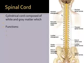
The Spinal Cord • Cylindrical in shape. • Occupies upper 2/3rd of the vertebral canal. • Begins superiorly at the Foramen magnum (Continuous with the Medulla Oblongata of Brain) • Ends inferiorly in adults at the Lower border of 1st Lumbar vertebra. • Surrounded by meninges & CSF.
The Spinal Cord • Fusiformly enlarged: • Cervical enlargement • Lumbar enlargement • Inferiorly tapers off into Conus Medullaris.
The Spinal Cord • Attached to coccyx by a Pia Mater extension (filum terminale).
The Spinal Cord Denticulate Ligament
Spinal Cord (Grooves or Sulcus) • Anterior median fissure • Posterior median sulcus
Spinal Cord (Internal Structure) • Gray matter in the center (H-shaped). • Posterior or Dorsal horns • Anterior or Ventral horns. • Lateral horn is only present in the thoracic, lumbar and sacral Regions. • The intermediate zone pierced by Central Canal.
Spinal Cord (Internal Structure) • White matter divided into: • Ventral Column or Funiculus • Dorsal Column or Funiculus • Lateral Column or Funiculus
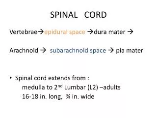
Meninges of Spinal Cord • Are continuous with the cranial meninges. • Dura mater • Arachnoid mater • Pia mater
Meninges of Spinal Cord • Epidural space: • Lies between vertebral column and dura mater • Contains blood vessels, areolar connective tissue & fat. • Subdural space: • Lies between the dura mater and arachnoid mater • Contains serous fluid. • Subarachnoid space: • Lies between arachnoid mater and Pia mater. • Contains cerebrospinal fluid (CSF) and blood vessels
The Spinal Cord • 8 Cervical nerves (C1-C8) • 12 Thoracic nerves (T1-T12) • 5 Lumbar nerves (L1–L5) • 5 Sacral nerves (S1–S5), • 1 Coccygeal nerve
The Spinal Cord • White matter • Myelinated axons • Divided into three columns (funiculi) • Ventral • Dorsal • lateral • Commissures: Connections between left and right halves • Gray with central canal in the center • White • Roots • Spinal nerves rootlets (dorsal and ventral roots). • Dorsal and ventral roots merge to form the spinal nerve.
Gray Matter • Consists of nerve cell bodies and their processes, neuroglia, and blood vessels • The nerve cells are multipolar and are of three main categories: • Sensory neurons: Receive impulses from the periphery of the body and whose axons constitute the ascending fasciculi of the white matter, are located in the dorsal horns • Motor neurons, which transmit impulses to the skeletal muscles, are located in the ventral horns (similar neurons in the lateral horn are the preganglionic neurons of the autonomic system) • Interneurons (Connector neurons) : linking sensory and motor neurons, at the same or different levels, which form spinal reflex arcs.