Download

1 / 81
820 likes | 1.24k Vues
The Spinal Cord and Spinal Nerves. Dr. Michael P. Gillespie. Medical Terminology. Kyphosis – exaggeration of the thoracic curve Lordosis – an exaggeration of the lumbar curve or cervical curve Lumbar spine stenosis – narrowing of the spinal canal

E N D
The Spinal Cord and Spinal Nerves Dr. Michael P. Gillespie
Medical Terminology • Kyphosis – exaggeration of the thoracic curve • Lordosis – an exaggeration of the lumbar curve or cervical curve • Lumbar spine stenosis – narrowing of the spinal canal • Scoliosis – lateral bending of the vertebral column
Protective Structures • Bony vertebrae • Meninges • Cerebrospinal fluid (produced in the brain)
Vertebral Column • The spinal cord is located within the vertebral canal of the vertebral column. • The vertebral foramina form the canal. • The vertebrae form a shelter for the cord. • The vertebral ligaments, meninges and CSF also provide protection.
Functions of the Spinal Cord and Spinal Nerves • White matter – contains the sensory and motor tracts (“highways”). • Gray matter – site for integration (summing) of action potentials. • Spinal nerves – connect the CNS to sensory receptors, muscles, and glands.
Meninges • Connective tissue coverings that encircle the spinal cord and brain. • Spinal meninges. • Cranial meninges.
Meninges • 3 spinal meninges. • Dura mater (most superficial). • Epidural space – between dura mater and wall of vertebral canal. • Arachnoid mater (middle layer) – spider web arrangement. • Subdural space – between dura and arachnoid. • Pia mater (innermost layer). • Subarachnoid space – between arachnoid and pia – contains CSF. • Denticulate ligaments – extend from pia and fuse with arachnoid.
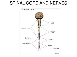
External Anatomy of the Spinal Cord • Cervical enlargement – nerves to and from the upper limbs • Lumbar enlargement – nerves to and from the lower limbs
External Anatomy of the Spinal Cord • Conus medullaris – the spinal cord tapers to a conical portion • Filum terminale – an extension of the pia mater that anchors the spinal cord to the coccyx
External Anatomy of the Spinal Cord • Cauda equina “horse’s tail” • Spinal nerves – paths of communication between the cord and the nerves innervating specific regions of the body • Posterior (dorsal) root • Sensory nerve axons • Posterior (dorsal) root ganglion – swelling – cell bodies • Anterior (ventral) root • Motor nerve axons
Spinal Tap • Spinal tap (lumbar puncture). • Local anesthetic is given and a long needle is inserted into the subarachnoid space. • Uses. • Withdraw CSF for diagnosis. • Introduce antiobiotics, contrast media, anesthetics. • Introduce chemotherapy. • Measure CSF pressure.
Internal Anatomy of the Spinal Cord • Anterior median fissure. • Posterior median sulcus.
Internal Anatomy of the Spinal Cord • Gray commissure – form the crossbar of the “H.” • Anterior (ventral) gray horns – cell bodies of somatic motor neurons and motor nuclei. • Posterior (dorsal) gray horns - cell bodies of somatic and autonomic sensory nuclei. • Lateral gray horns – cell bodies of autonomic motor neurons that regulate smooth muscle, cardiac muscle and glands. • Central canal – in the center of the gray commissure.
Internal Anatomy of the Spinal Cord • White columns. • Sensory (ascending) tracts. • Motor (descending) tracts.
Spinal Cord Physiology • 2 principle functions. • Nerve impulse propagation – white matter tracts. • Sensory impulses flow toward the brain. • Motor impulses flow from the brain. • Information integration – gray matter.
Sensory & Motor Tracts • The name of the tract often indicates its position in the white matter and where it begins and ends.
Sensory Tracts • Lateral and anterior spinothalamic tracts. • Convey impulses for pain, warmth, tickling, itching, deep pressure, and a crude sense of touch (poorly localized). • Posterior columns. • Convey impulses for proprioception, discriminative touch, 2 point discrimination, light pressure sensations, and vibrations.
Motor Tracts • Direct pathways – convey precise voluntary movements. • Lateral corticospinal. • Anterior corticospinal. • Corticobulbar.
Motor Tracts • Indirect pathways – govern automatic movements (I.E. Reflexes). • Rubrospinal. • Tectospinal. • Vestibulospinal.
Reflexes and Reflex Arcs • Reflex – a fast, unplanned sequence of actions that occurs in response to a particular stimulus. • Location of integration. • Spinal reflex. • Cranial reflex – integration in brain stem. • Types of reflexes. • Somatic reflexes – contraction of skeletal muscles. • Autonomic (visceral) reflexes – responses of smooth muscle, cardiac muscle, and glands.
Reflex Arc • Reflex arc (reflex circuit) - the pathway followed by nerve impulses.
Five Functional Components of a Reflex Arc • Sensory receptor. • Distal end of a sensory neuron. • Responds to a stimulus. • Sensory neuron. • Nerves terminate in the brain stem or spinal cord.
Five Functional Components of a Reflex Arc • Integrating center. • Monosynaptic reflex arc - A synapse between a sensory neuron and a motor neuron. • Polysynaptic reflex arc – one or more interneurons and a motor neuron.
Five Functional Components of a Reflex Arc • Motor neuron. • Effector. • The part of the body that responds to the motor nerve impulse. • Somatic reflex – the effector is a skeletal muscle. • Autonomic reflex – the effector is smooth muscle, cardiac muscle or a gland.
Reflexes • Reflexes are normally predictable. • They can provide information about the health of the nervous system.
Reflexes • Damage or disease anywhere along the reflex arc can cause the reflex to be absent or abnormal. • Somatic reflexes can be tested by tapping or stroking the body surface. • Autonomic reflexes cannot be tested easily because the visceral receptors are deep inside the body.
Four Important Somatic Spinal Reflexes • Stretch reflex • Tendon reflex • Flexor (withdrawal) reflex • Crossed (extensor) reflex
Stretch Reflex • A stretch reflex causes contraction of a skeletal muscle in response to stretching of the muscle. • Monosynaptic reflex arc. • Ipsilateral reflex. • This reflex helps avert injury by preventing overstretching of a muscle. • Reciprocal inhibition – when the stretched muscle contracts, the antagonistic muscle(s) relax.
Tendon Reflex • The tendon reflex controls muscle tension by causing muscle relaxation before muscle forces become so great that they tear tendons. • Ipsilateral reflex. • Sensory receptors – tendon (Golgi tendon) organs.
Flexor (Withdrawal Reflex) • Causes withdrawal from a painful stimulus. • This reflex causes contraction of the flexor muscles with causes withdrawal from a painful stimulus. • Ipsilateral reflex. • Several motor units at different levels of the spinal cord are recruited – intersegmental reflex arc. • Reciprocal innervation occurs.
Crossed Extensor Reflex • Helps you maintain balance. • Contralateral reflex arc. • Reciprocal innervation occurs.