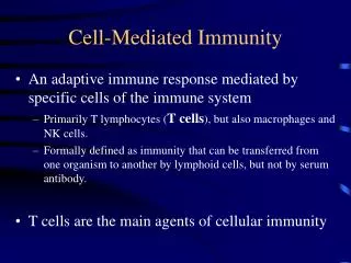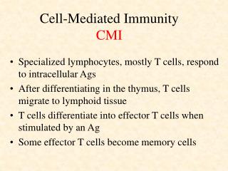

Cell Mediated Immunity
E N D
Presentation Transcript
1. Cell Mediated Immunity
2. T Lymphocytes
3. T-Cell Activation
4. T-Cell Activation
5. T-Cell Subpopulation
6. T-Cell Activation
7.
CD2 and Icam binding allow for specific antigen recognition to take place.
CD2 and Icam binding allow for specific antigen recognition to take place.
10. Ag recognition occurs in association with recognition of MHC II proteins.
It has been demonstrated that CD4 is a ligand for MHC class 2 molecules. The main function of CD4 appears to be an adhesion molecule to increase the avidity of binding between the Tcell and the target . It acts as a stabilizing bond and causes activation of T cells.Ag recognition occurs in association with recognition of MHC II proteins.
It has been demonstrated that CD4 is a ligand for MHC class 2 molecules. The main function of CD4 appears to be an adhesion molecule to increase the avidity of binding between the Tcell and the target . It acts as a stabilizing bond and causes activation of T cells.
11. Activated T cells must be able to divide quickly and produce large numbers of its progeny. This is dependent on a growth factor known as IL2.
CD28 signaling causes the production of IL2. At the same time the activated T-cell produces large amounts of IL2 receptors. This is known as autocrine stimulation. This results in an �army� of cells that can respond to a single specific antigen without additional stimulation.
In addition of effector cells, memory T cells are also stimulated. These are long lived cells that when antigen activated will respond rapidly to the stimulus by the same antigen ( I.e. small pox vaccin)Activated T cells must be able to divide quickly and produce large numbers of its progeny. This is dependent on a growth factor known as IL2.
CD28 signaling causes the production of IL2. At the same time the activated T-cell produces large amounts of IL2 receptors. This is known as autocrine stimulation. This results in an �army� of cells that can respond to a single specific antigen without additional stimulation.
In addition of effector cells, memory T cells are also stimulated. These are long lived cells that when antigen activated will respond rapidly to the stimulus by the same antigen ( I.e. small pox vaccin)
12. Triggering of IL1 Antigen specific T helper cells also influence APC. APC release IL-1 that act on T-cells to promote IL2 production and increases the number of receptors in T helper cells.Antigen specific T helper cells also influence APC. APC release IL-1 that act on T-cells to promote IL2 production and increases the number of receptors in T helper cells.
13. T- Helper Subsets
14. Stimulation of B-Cells Activated T cells must find the target B cells containing the same antigen in conjuction with MHC2. The binding between T cell and B cell is not initially antigen specific and occurs by adhesion of CD2 and LFAActivated T cells must find the target B cells containing the same antigen in conjuction with MHC2. The binding between T cell and B cell is not initially antigen specific and occurs by adhesion of CD2 and LFA
15. Memory B cells are essentially a proliferating clone that does not differentiate into Plasma cells. These cell can survive for years within lymphoid follicles. If subsequently activated they can produce more memory effector cells (Booster in vaccins) It is the mimory cells that are responsible for the the primary and secondary responses.Memory B cells are essentially a proliferating clone that does not differentiate into Plasma cells. These cell can survive for years within lymphoid follicles. If subsequently activated they can produce more memory effector cells (Booster in vaccins) It is the mimory cells that are responsible for the the primary and secondary responses.
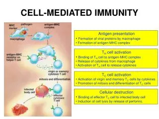
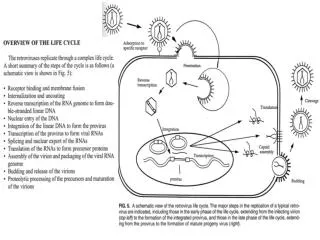
17. Activation of Cytotoxic T-Cells Overview of Cell-Mediated Immunity
The human immune system is made up of many different kinds of cells that are
responsible for eliminating harmful invaders, such as viruses or cancer, from the body.
One cell of the immune system, the T cell (or T lymphocyte), plays the central role in
orchestrating most immune responses.
T cells become activated when they recognize antigens as foreign to the body (Figure
1). This occurs when antigens are picked up and processed into peptides which bind
to the major histocompatibility complex (MHC) of an antigen-presenting cell (APC;
note: dendritic cells are the most well-known APCs). These peptide/MHC complexes
are translocated to the surface of an APC, where they bind to the T cell receptor (TCR)
on a T lymphocyte. The TCR is closely associated with the CD3 signaling complex
(TCR/CD3 complex) on a T lymphocyte.
The primary signal for activating a T cell takes place when the TCR binds to a
peptide/MHC complex on an APC (Figure 1). Each individual T cell only expresses a
single TCR capable of recognizing a specific antigen. However, the many billions of T
cells found in the human body express millions of different TCRs, thereby enabling
recognition of millions of distinct antigens. Only a specific T cell that recognizes a
particular antigen will become activated in a normal immune response.
Xcyte Therapies' scientific founders discovered that APCs must also deliver a second
signal in order to activate T cells. This signal occurs when co-stimulatory ligands on
APCs bind to co-stimulatory CD28 receptors on T cells (Figures 1). Activation of T
cells takes place when the major histocompatability complex (MHC) and other specific
receptors on APCs bind to the TCR/CD3 complex and CD28 receptor, respectively.
These activated T cells are exquisitely sensitive to further stimulation and also secrete
a variety of chemical messengers called cytokines. This process further augments the
immune response both by driving continued activation and proliferation of T cells and
recruiting and stimulating other cells of the immune system. This cascade of events
ultimately leads to destruction of pathogens such as tumor cells and viruses.
Overview of Cell-Mediated Immunity
The human immune system is made up of many different kinds of cells that are
responsible for eliminating harmful invaders, such as viruses or cancer, from the body.
One cell of the immune system, the T cell (or T lymphocyte), plays the central role in
orchestrating most immune responses.
T cells become activated when they recognize antigens as foreign to the body (Figure
1). This occurs when antigens are picked up and processed into peptides which bind
to the major histocompatibility complex (MHC) of an antigen-presenting cell (APC;
note: dendritic cells are the most well-known APCs). These peptide/MHC complexes
are translocated to the surface of an APC, where they bind to the T cell receptor (TCR)
on a T lymphocyte. The TCR is closely associated with the CD3 signaling complex
(TCR/CD3 complex) on a T lymphocyte.
The primary signal for activating a T cell takes place when the TCR binds to a
peptide/MHC complex on an APC (Figure 1). Each individual T cell only expresses a
single TCR capable of recognizing a specific antigen. However, the many billions of T
cells found in the human body express millions of different TCRs, thereby enabling
recognition of millions of distinct antigens. Only a specific T cell that recognizes a
particular antigen will become activated in a normal immune response.
Xcyte Therapies' scientific founders discovered that APCs must also deliver a second
signal in order to activate T cells. This signal occurs when co-stimulatory ligands on
APCs bind to co-stimulatory CD28 receptors on T cells (Figures 1). Activation of T
cells takes place when the major histocompatability complex (MHC) and other specific
receptors on APCs bind to the TCR/CD3 complex and CD28 receptor, respectively.
These activated T cells are exquisitely sensitive to further stimulation and also secrete
a variety of chemical messengers called cytokines. This process further augments the
immune response both by driving continued activation and proliferation of T cells and
recruiting and stimulating other cells of the immune system. This cascade of events
ultimately leads to destruction of pathogens such as tumor cells and viruses.
18. Rationale for Developing Xcellerate
Inadequate or impaired antigen presentation and/or lack of primary or co-stimulatory
immune activation signals appears to play a major role in the failure of the immune
system to detect and eradicate cancer cells. Therefore, strategies that boost or
restore these immune responsive elements of a patient's T cells might overcome these
deficits. Since the primary mechanism of T cell activation requires co-stimulation of
both the TCR/CD3 complex and the CD28 receptor on the T cell surface, mimicry of
this event in a defective immune system could provide a strong stimulus to overcome
the impairment. In this respect, antibodies directed against CD3 have been shown to
be capable of providing an activation signal through the CD3 component of the
TCR/CD3 complex. In addition, specific antibodies directed against CD28 have been
shown to enhance T cell proliferation and cytokine secretion in response to a variety of
stimuli.
The combined use of anti-CD3 and anti-CD28 antibodies can, therefore, be used to
activate T cells and initiate and/or enhance immune responses (Figures 2 and 3). Rationale for Developing Xcellerate
Inadequate or impaired antigen presentation and/or lack of primary or co-stimulatory
immune activation signals appears to play a major role in the failure of the immune
system to detect and eradicate cancer cells. Therefore, strategies that boost or
restore these immune responsive elements of a patient's T cells might overcome these
deficits. Since the primary mechanism of T cell activation requires co-stimulation of
both the TCR/CD3 complex and the CD28 receptor on the T cell surface, mimicry of
this event in a defective immune system could provide a strong stimulus to overcome
the impairment. In this respect, antibodies directed against CD3 have been shown to
be capable of providing an activation signal through the CD3 component of the
TCR/CD3 complex. In addition, specific antibodies directed against CD28 have been
shown to enhance T cell proliferation and cytokine secretion in response to a variety of
stimuli.
The combined use of anti-CD3 and anti-CD28 antibodies can, therefore, be used to
activate T cells and initiate and/or enhance immune responses (Figures 2 and 3).
22. Cell death occurs within 10 minutes to 3 hours.
TNF-B has life of only minutes
TNF B acts only locally
Effector cell does not die and is able to seek other targetsCell death occurs within 10 minutes to 3 hours.
TNF-B has life of only minutes
TNF B acts only locally
Effector cell does not die and is able to seek other targets
24. in order to mimic co-stimulation, Xcyte Therapies utilizes super-paramagnetic
microbeads that approximate the size and shape of APCs. By covalently attaching
anti-CD3 and anti-CD28 monoclonal antibodies to these microbeads, artificial APCs
are created that can be used to activate T cells (Figure 3). Furthermore, the
super-paramagnetic nature of the microbeads makes it feasible to remove them with a magnetic device after completion of the T cell activation procedure.
in order to mimic co-stimulation, Xcyte Therapies utilizes super-paramagnetic
microbeads that approximate the size and shape of APCs. By covalently attaching
anti-CD3 and anti-CD28 monoclonal antibodies to these microbeads, artificial APCs
are created that can be used to activate T cells (Figure 3). Furthermore, the
super-paramagnetic nature of the microbeads makes it feasible to remove them with a magnetic device after completion of the T cell activation procedure.
25. Role of Cytokines : Interleukin-1
26. Role of Cytokines : Interleukin-2
27. Role of Cytokines : Interleukin-3
31. Role of Cytokines : Interleukin-4
34. Lymphocyte Function: Blast Formation
36. Applied Immunology: Photopheresis
37. Photopheresis