Download
1 / 24
240 likes | 254 Vues
This article examines the role of mitochondrial dysfunction in Parkinson's disease (PD). It discusses the defects in the ubiquitin-proteasome system (UPS) and the accumulation of misfolded proteins, as well as genetic mutations in a-synuclein, Parkin, and PINK-1. The article also explores the impact of oxidative stress and mitochondrial DNA deletions on cell death in PD.

E N D
Background • defects in the capacity of the ubiquitin-proteasome system (UPS) to degrade unwanted proteins is considered to play a role in the pathogenesis of PD • Synthetic PSI (Z-lle-Glu(OtBu)-Ala-Leu-al), an inhibitor of the proteasome, selectively impairs the enzyme in a reversible manner • A rat model of PD based on inhibition of proteasomal function and UPS-mediated protein degradation has been recently proposed (McNaught KSP, et al. Ann Neurol 2004;56:149–162).
Pathology • Loss of SN pigmented dopamine neurons • Lewy bodies • Lewy neurites-multiple brain regions • Lewy bodies stain with antibodies to alpha synuclein, ubiquitin, others • Also present in autonomic and submucosal ganglia • Clear that PD is more than just a disorder of dopamine deficiency, but that SN cells for an unknown reason are even more sensitive to the stresses of the pathological abn than other parts of the brain
Mito dysfunction • In PD, SN neurons accumulate mito DNA deletions at an abn rate-suggests that oxidative stress is occurring. • Impaired cell respiration results from mito DNA deficiency that causes respiratory chain deficiency • A mutation in the gene for mito DNA polymerase assoc. with accumulation in deletions of mito DNA, SN loss, early PD • Common feature of PD is evidence of Complex 1 deficiency • Complex 1 also affected by rotenone and MPTP • When rotenone given chronically to rodents, it causes complex 1 deficiency, dopaminergic cell loss in SN
Mito dysfunction • 6-hydroxydopamine and paraquat cause oxidative stress, mimic mito toxicity seen with MPTP • Findings led to trials of coenzyme Q, vit E, creatine, all anti-oxidant and pro-mitochondrial compounds
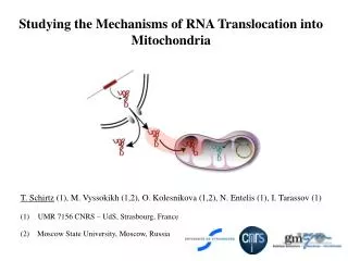
Mitochondria in PD • Contributions to understanding the pathogenesis of PD by familial inherited forms of PD
Genetic mutations-a-synuclein • First to be identified was a-synuclein • Point mutations caused familial PD, rare AD form • Mice lacking gene for a-synuclein show resistance to MPTP-induced dopaminergic toxicity • In Lewy bodies it is present in aggregated form in insoluble filaments that are hyperphosphorylated and ubiquitinated • It is likely that misfolded a-synuclein is toxic to neurons • Factors that increase aggregation of a-synuclein are genetic mutations, proteasome and mitochondrial dysfunction, oxidative stress, phosphorylation. • Likely involved in synaptic vesicle function
Genetic mutations-Parkin • Mutations in gene for Parkin cause aut. Recessive form of PD • Most common genetic cause-50% with family history • Parkin is an E3 ligase-participates in addition of ubiquitin molecules to target proteins, marking them for degradation by the proteasome • Loss of parkin function therefore leads to an inability to break down toxic substances with subsequent neuronal dysfunction and cell death. • Parkin substrates p38/JTV and FBP-1 accumulate in sporadic cases of PD and in Parkin K/O mice • Role of ubiquitination in development of PD is a promising field of study
PINK-1 • Mutations in this gene encoding PTEN (Phosphatase and tensin homologue)-induced putative kinase 1(PINK-1) cause aut. recessive PD. • Mitochondrial protein kinase, substrates unknown • Targets to mitochondria • K/O in Drosophila assoc. with mitochondrial dysfunction, reduced respiratory chain activity, reduced mito DNA, reduced ATP content of tissues and increased propensity to apoptosis of affected cells such as muscle • Parkin over-expression rescues the loss of function phenotype of PINK-1 K/O in Drosophila, Parkin downstream of PINK-1-links mitochondria to proteasome • Patients with genetic mutations in Parkin or PINK-1 are clinically indistinguishable
18 rats were injected with 6.0mg/kg (SC) PSI (Peptides International) dissolved in 10% dimethyl sulfoxide (DMSO) and 14 rats with 10% DMSO (SC) as a control. In each case, six injections were made over the course of 2 weeks (Mon, Wed, Fri, Mon, Wed, Fri).
calcium uniporter innermembrane Ca2+ outer membrane sodium-calcium exchanger -160 mV Ca2+ Na2+ permeability transition pore Ca2+ Inner membrane channels
In a previous study [Bonanni L, et al. J Neurosci. 2006 Jun 21;26(25):6851-62] we found a large mitochondrial channel activity to be present in a rat model of global ischemia and it was associated with appearance of the pro-apoptotic N-terminal proteolytic cleavage fragment, DN-BCL-xL, and protease activity in post-ischemic mitochondria, consistent with a role for cleaved BCL-xL in channel formation.
PSI-treated mitochondria exhibit an opening of large/intermediate conductance channels. Recordings were made from organelle-attached patches on the membranes of isolated PSI-treated and control diencephalic mitochondria. A. Sample recordings at -100 mV in PSI-treated (left) and control (right) diencephalic mitochondria.
Assay of mitochondrial function • Can protein aggregates produce or aggravate mitochondrial dysfunction? • Can the mito dysfunction cause neuronal death of sensitive neurons?
Neuronaldeath caspase-3 caspase-9 BAX BCL-2 proteins induce apoptosis by releasing cytochrome c from mitochondria Intermembranespace inner membrane outer membrane VDAC BAX cytochrome c
The mitochondrial permeability transition pore is a double membrane-spanning ion channel The mitochondrial permeability transition pore is a double membrane-spanning ion channel BAD VDAC/BCL-xL VDAC mPTP mPTP Outer mitochondrial membrane Inner mitochondrial membrane ANT CyD Cytochrome c Ca2+ or Zn2+ Messenger
Measuring death channel activity with the mitochondrial recording technique
KNS-760704 [R(+)Pramipexole] • Optical enantiomer of Mirapex without the dopaminergic component • Mirapex has utility for neuroprotection in PD (and other diseases) that is limited by its dopaminergic side-effects. • Both compounds are neuroprotective by an unknown mechanism-likely mitochondrial, independent of dopamine receptor affinity.
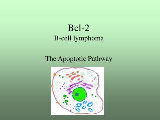
ABT-737 is a specific inhibitor of Bcl-xL, Bcl-2, and Bcl-w • Rationally designed BAD mimetic binds to hydrophobic cleft of BCL-xL • Displaces pro-apoptotic BH3-domain proteins to initiate cell death pathway in non-neuronal cells • Stage II clinical trials as anti-cancer drug Oltersdorf et al., 2005
ABT-737 inhibits cell death after ischemia in hippocampal CA1 neurons