Cell Death Pabio 552 5/11/06
560 likes | 1.28k Vues
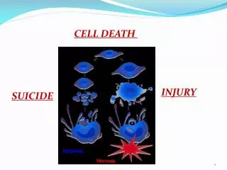
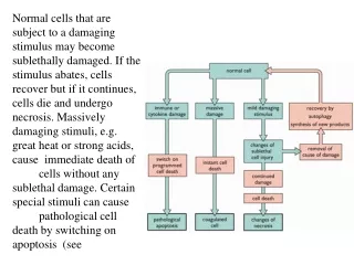
Cell Death Pabio 552 5/11/06. Classical View of Cell Death: Apoptosis vs Necrosis . Murder?. Suicide?. www.imm.ki.se/ sft/bilder/Image1.jpg. Types of cell death.

Cell Death Pabio 552 5/11/06
E N D
Presentation Transcript
Classical View of Cell Death: Apoptosis vs Necrosis Murder? Suicide? www.imm.ki.se/ sft/bilder/Image1.jpg
Types of cell death Necrosis – non-apoptotic accidental cell death (common definition) <or> morphology seen after a cell has already died (pathology; has nothing to do with biochemistry of how the cell died) Autophagy – degradation of cellular components within dying cells in an autophagic vacuole; begins with sequestration of cytoplasmic material within phagosomes, under control of GTPases and phosphatidylinositol kinases Oncosis – prelethal pathway leading to cell death; accompanied by cellular swelling, organelle swelling, blebbing, and increased membrane permeability; oncotic cells proceed to necrosis with lysis and spillage of contents before being recognized by phagocytosis; inflammation results Pyroptosis – induced by infection with Salmonella and Shigella; inherently proinflammatory; dependent on caspase 1 Apoptosis…..
Apoptosis An active programmed process of autonomous cellular dismantling that avoids eliciting inflammation Characterized by: - exposure of phosphotidyl serine on cell surface - cytoplasm shrinkage - membrane blebbing - chromatin condensation - cleavage of DNA at internucleosome site - caspase mediated - corpse clearance via phagocytosis Fink et al Inf. Imm. Apr.2005: 1907-16
Biological Roles for Apoptosis Development Metamorphosis Regulation of cell number in tissues (homeostasis and tumorigenesis) Immune defense (cytotoxic T cell activity) Develoment of B and T cells via negative selection Disease: Cancer, autoimmunity, infectious disease etc.
Caspases Cysteine-dependent aspartate specific proteases Have a cysteine at the active site Cleave target just after aspartic acid residues Substrate specificity is determined by the 4 residues upstream of cleavage site Exist in cytosol as single chain proenzymes (procaspases) which are activated when cleaved by other caspases Once activated, cleave other caspases – results in proteolytic cascade Also cleave key proteins in the cell, causing the characteristic morphology and biochemistry of apoptosis.
Caspases… Procaspases - Contain N terminal pro-domain followed by region that forms a 2 subunit catalytic effector domain Prodomain: for prot-prot interactions; allows it to bind upstream regulators and effector proteins; examples include: DED – death effector domain (e.g. caspase 8) CARD – caspase activation and recruitment domain (e.g. caspase 9) Active caspases – heterotetramers composed of two large and two small subunits with two active sites per molecule Nature407, 770-776 (12 October 2000)
Caspases… Two types: - those related to caspase 1 (Caspases 1, 4, 5, 13, and 14); role in cytokine processing during inflammation - those involved in apoptosis (Caspases 2, 3, 6, 7, 8, 9 and 10) Initiators – activate downstream effector caspases to initate activation cascades Effectors - cleave target proteins resulting in morphological and biochemical markers of apoptosis
Effector Caspases Activated effector caspases cleave target proteins: Nuclear Lamins – scaffold proteins of nuclear envelope; leads to nuclear shrinkage and fragmentation Cytoskeleton proteins – e.g.: Fodrin; leads to loss of cell shape and membrane blebbing; Gelsolin (an actin depolymerizing enzyme); cleaved by caspase 3; role in cell morphology during apoptosis (blebbing etc.) ICAD (inhibitor of Caspase Activated Dnase) – DNA now cut up by CAD Components of focal adhesion complex; leads to detachment of apoptotic cells from other cells Other caspase dependent features: Cleavage of PAK2 (member of p21-activated kinase family); results in formation of apoptotic bodies and other signaling cascades Exposure of phosphatidylserine on outer membrane; probably due to down-regulation of phospholipid translocase activity and/or activation of lipid scramblase
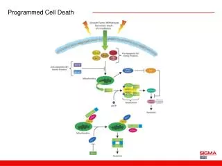
Caspase Pathways Intrinsic pathway – mitochondria mediated; caspase 9 Extrinsic pathway – involves death receptors (TNFreceptor, Fas); caspase 8 Converge to active executioner caspases 3 and 7 Hail et al. Apoptosis (2006)
Intrinsic Pathway Usually initiated by cellular stress (UV, cytotoxic drugs etc.) usually causing alterations in mitochondria membrane potential (MMP). Mitochondria-dependent: mitochondria sequester pro-apoptotic proteins, e.g. cytochrome C. Regulated by Bcl-2 family member of proteins – regulate the release of pro-apoptotic factors from mitochondria. Changes in MMP free cytochrome c from intermembrane space out into the cytosol Cytochrome c then combines with dATP, APAF-1 (apoptotic protease activating factor-1), and caspase 9 to form a catalytic complex called the apoptosome (Caspase 9 co-factor is APAF-1 which must be bound by cytochrome c to drive Caspase 9 into its active conformation) Apoptosome activates caspases 3 and 7 (effector caspases) Effector caspases Nature407, 770-776 (12 October 2000)
Regulation of intrinsic pathway Bcl-2 family of proteins (gene orig. isolated from a B-cell lymphoma): Pro apoptotic effects: e.g. Bax (indirectly regulated by tumor repressor P53), Bad, and Bak Anti apoptotic effects: e.g. Bcl-2, Bcl-XL These proteins may be localized to mitochondria intermembrane or targeted to the organelle in response to stimulus Regulate release of pro-apoptotic factors from mitochondria, especially cytochrome c. Also release Smac (second mitochondria-derived activator of caspases) and DIABLO (direct IAP-binding protein with low pI) which bind to IAPs (Inhibioros of Apoptosis proteins) Battle between pro and anti levels to determine cell’s response to apoptotic stimuli such as ROS (reactive oxygen species), Ca++, radiation etc. Also released from mitochondria is AIF – apoptosis inducing factor; once released, it translocates to nucleus to induce chromatin condensation and DNA fragmentation
How do proteins get released from mitochondria? Several hypotheses for how Bcl-2 proteins regulate release of cytochrome C: By forming channels in mitochondria membrane By interacting with other proteins to form channels By inducing rupture of the mitochondrial membrane By oligomerizing to form a weakly selective ion channel. BcL-2 family proteins regulate the release of apoptogenic cytochrome c by the mitochondrial channel VDAC (voltage dependent anion channel) Nature 399, 483-487 (1999))
Extrinsic Pathway Initiated by death receptors: Start with ligand binding, clustering or aggregation of death receptors Cytoplasmic tails of death receptor bind adaptor proteins Adaptor proteins recruit and activate procaspase 8 to yield caspase 8 (active) Caspase 8 triggers downstream effector caspases, such as caspase 3 Activation of death receptors may also activate intrinsic pathway Death Receptors: TNFR family Fas (CD95) Death Ligands: TNF CD95L(FasL) – only expressed by activated T cells Nature407, 770-776 (12 October 2000)
Fas – Fas ligand system Fas Prototypical cell death receptor of the TNF receptor superfamily No intrinsic enzymatic activity Signals via adaptor proteins Fas ligand (FasL) Only expressed by activated T cells Transmembrane TNF-like protein When the TCR (T cell receptor) of an antigen specific CTL (cytotoxic T lymphocyte) binds antigen on MHC I, the expression of FasL is induced on the T cell FasL binds Fas (present on most cells of body) that is on the presenting cell to induce death of that cell Cytoplasmic tail of Fas binds its adaptor protein FADD (Fas Associated Death Domain protein) Fas-FADD complex binds to and activates caspase 8 Initiates lethal proteolytic cascade
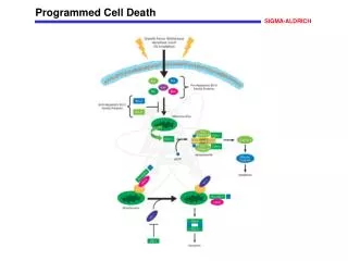
Extracellular ligands inducing apoptosis via diverse signal transduction pathways
Corpse clearing A defining point of apoptosis is to clear the dying cell before it can release inflammatory molecules Macrophages ingesting apoptotic cells release anti-inflammatory and immunosuppressive cytokine transforming growth factor-beta1 (TGF-β1) Macrophages ingesting necrotic cells will release pro-inflammatory mediators Cells undergoing apoptosis show changes in surface of plasma membrane. These “eat me” signals are recognized by phagocytes Exposure of phosphatidyl serine (PS) – PS receptor is expressed on phagocytes Change in cell surface sugars – detected by lectins on phagocyte cell Sites that bind “bridging molecules” e.g. C1q – C1q deficiency leads to impaired phagocytosis of apoptotic cells ICAM-3 – binds alternate receptor on macrophages; possibly CD14 Macrophages thought to “tether” dying cells by using CD14 or beta integrin before engaging receptors that drive apoptosis
Jurkat T-cell targets were labelled with 5-(and 6)-carboxytetramethylrhodamine succinimidyl ester and irradiated to induce apoptosis. The macrophages were stained with fluorescein isothyocyanate-conjugated phalloidin to identify actin filaments. Nature (407) pp 784-788.
Cell survival Many signaling pathways exist to promote cell survival Often dependent on growth factors or cell-cell interactions (survival factors) Example: PI 3-Kinase initiated signaling cascase PI3K is activated by tyrosine kinases or G coupled protein receptors PI3K phosphorylates PIP2 to form PIP3 which activates the serine threonine kinase Akt Akt phosphorylates many proteins involved in regulation of apoptosis: Bad (induces release of cytochrome c from mitochondria); phosphorylation of Bad creates binding sites for proteins to sequester Bad in the cytosol to keep it from going to the mitochondrial membrane Caspase -9 Transcription factors e.g. NFkB GSK-3; affects metoblism and protein synthesis Other signaling pathways include: Ras/Raf/MAP kinase
PI 3-kinase/Akt signaling cascade © 2000 by Geoffrey M. Cooper
Physiological TNF family (TNF, FasL) TGF-beta Neurotransmitters Growth factor withdrawal Loss of matrix attachment Sustained rise in Calcium Glucocorticoids Damage related Heat Shock Viral infection Bacterial toxins Oncogenes Tumor supressors (p53) Cytolytic T cells Oxidants Free radicals Nutrient deprivation UV radiation Gamma radiation Inducers of apoptosis Toxins Some chemotherapeutic drugs Ethanol Beta-amyloid peptide
Physiological Growth factors ECM CD40L Neutral a.a. Zinc Estrogen Androgens Viral gene products Adenovirus E1B Baculovirus p35 Cowpox virus crmA Epstein-Barr virus BHFR1, LMP1 African swinve fever virus LMW5-HL Herpesvirus gamma 1 34.5 Inhibitors of apoptosis Pharmacological agents Calpain inhibitors Cysteine protease inhibitors Tumor promoters e.g. phenobarbital
Assays for detecting apoptosis Histological stains to look at condensed chromatin DNA fragmentation assessed by gel electrophoresis - CAD (caspase activated DNase) is present in cells bound to its inhibitor (ICAD - inhibtor CAD); activation of CAD occurs when ICAD is cleaved; CAD cleaves genomic DNA to generate strands ~ 180 bp; forms “DNA ladder” used as a marker for an apoptosing cell Annexin V staining - stains phosphatidyl serine on outer membrane; when fluorescently conjugated, can be sorted by FACS TUNEL (terminal deoxynucleotidyl transferase-mediated dUTP nick end labeling) – uses activity of the terminal deoxynucleotidyl transferase enzyme to label the 3’ ends of DNA strand breaks which may then be identified by microscopy Caspase activation – can be demonstrated by Western using Abs against caspase substrates; also measured by using colorimetric and fluorometirc assays based on proteolysis of conjugated tetrapeptide substrates mimicking caspase cleavage sites Cytochrome C release – assays measure cytochrome c in mitochondria vs cytosol Alterations in mitochondrial membrane potential – various assays to look at this; involve labeling specific molecules that move across mitochondria membrane; aid in calculating membrane potential
Apoptosis and Pathogens: Theileria parva Tick transmitted intracellular apicomplexans Worldwide infection in mammals Major constraint in livestock development, especially in tropical regions
T. parva vs. B. taurus leukocyte T. parva prevents apoptosis in host cells Upon infection, get induced expression of NFkB How? Parasite recruits IKK complex to its cell surface, get oligomerization causing “proximity induced activation” IKK complex stimulation results in phosphorylation of inhibitory IkB NFkB is now free to translocate to the nucleus Get upregulation of anti-apoptotic proteins e.g. cFLIP Alberts ch-17
Apoptosis and Pathogens: Enterovirus 71 Enterovirus 71 Infection Induces Fas Ligand Expression and Apoptosis of Jurkat CellsJournal of Medical Virology 78:780–786 (2006) EV71 is a +RNA virus Transmission – fecal-oral Clincial features: meningitis, encephalitis, pulmonary edema, death Px: increased cytokine levels, significant decrease in T cells EV71 induced FasL expression in Jurkat cells and increases Jurkat cell apoptosis
TUNEL assay A and B: mock infection of Jurkat cells C and D: EV71 infected Jurkat cells TUNEL labeling in green Cell nuclei in red (propidium idodide stain)
DNA fragmentation assay Jurkat cells incubated with or without EV71 for 48 hours. DNA samples run on 2% agarose gel and stained with ethidium bromide
Let down the bars, O death! The tired flocks come in Whose bleating ceases to repeat, Whose wandering is done. Thine is the stillest night, Thine the securest fold; Too near thou art for seeking thee, Too tender to be told. Emily Dickinson
Inhibition of caspase activation and a requirement for NF-kB function in the Toxoplasma gondii-mediated blockade of host apoptosis T. Matthew Payne, Robert E. Molestina and Anthony P. Sinai
Infection routes of T. gondii Undercooked meat Oocysts Reproductive Host Asexual Host predation
Tachyzoite Intracellular Bradyzoite Extracellular