Hip Arthroplasty
550 likes | 1.13k Vues
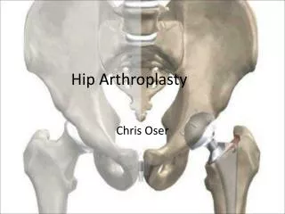
Hip Arthroplasty. Anatomy of Hip. Hip Joint. Ball and socket Ball is the femoral head Socket is Acetabulum Half sphere depression Lined with cartilage Horseshoe shape. Hip Joint. Femur Neck-shaft angle ~ 135 0 2/3 rd of head is covered with cartilage Head fits into acetabulum

Hip Arthroplasty
E N D
Presentation Transcript
Hip Joint • Ball and socket • Ball is the femoral head • Socket is Acetabulum • Half sphere depression • Lined with cartilage • Horseshoe shape
Hip Joint • Femur • Neck-shaft angle ~ 1350 • 2/3 rd of head is covered with cartilage • Head fits into acetabulum • Suction effect during dislocation
Hip OA • Cartilage gradually wear down • Femoral head and acetabulum grind on each other (bone-on-bone arthrosis)
Traumatic arthritis • Occurs following injury to hip • Direct trauma • damage to cartilage • Femoral neck fracture • Hip dislocation • Blood supply may be lost • Avascular necrosis
Rheumatoid arthritis • Body's immune system attacks synovium and cartilage • Joint arthrosis • Deformity • Stiffness • Women are more often affected than men
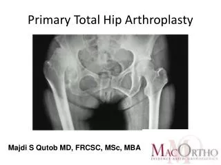
Plain X-rays • Loss of joint space • Subchondral sclerosis • Subchondral Cysts • Irregularity of joint surface • Subluxation
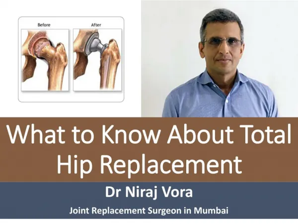
Objectives • Joint replacement • Femoral stem • IM Metal implant • Modular • Titanium stem and cobalt-chrome head • Acetabular cup • A low-wearing plastic insert • Press fit to acetabulum • Porous coated
Types of Implants • Implants may be • Cemented • Porous coated • Mesh of holes on implant surface • Secured as bone in grows
Acetabular component • Shell is made of metal • Plastic liner • Load bearing • Fits snugly inside shell
Femoral Stem • Made of metal • Usually titanium • Head • Diameter • 28, 32 mm • Material • Cobalt chrome • Ceramic
Surgical Procedure • An incision about eight inches long (dotted line) • Exposure hip joint • Anterior • Posterior
Removal of Femoral Head • Femoral head is dislocated from acetabulum • Neck cut • Femoral head is removed
Acetabulum Reaming • Acetabular cup is reamed into a hemisphere • Cartilage is removed
Reaming the Acetabulum • Lateral View
Inserting the Acetabular component • Acetabular shell • Porous coated • Press fit • Screws for stability • Cemented • A hard smooth plastic liner is inserted into metal shell
Reaming of Femoral Canal • Intramedullary canal finder • Manual insertion of a rod • Distal intramedullary reaming with a straight reamer • Rasping
Femoral Stem Insertion • Press fit • Cemented • Pressurization • Canal plug • Cement vacuum mix • Cement Gun
Femoral Head • A metallic head is attached to stem
Hip Reduction • Ball is reduced into acetabular liner • Soft tissue tension is tested • Leg length may be a problem
Animation of hip replacement • http://www.hipandkneesurgery.net/hip.html
Hybrid Fixation • Acetabular cup • Press fit • Femoral stem • Cemented
Care after Surgery • A suction drain • May be used for 1-2 days after surgery • Intravenous fluids & antibiotics • Pain medication • Elastic stockings, compression stockings and blood thinners • To decrease chances of blood clots • For first 6-8 weeks • Low sitting may cause dislocation
Care after Surgery • Physical therapy • Getting in and out of bed • Standing and walking • Crutches or a walker • Discharge from hospital • Usually in 3-5 days • Continued PT, OT
Complications • Thrombophlebitis • Blood clots within deep veins • Swelling of leg • Become warm to touch • Painful • May lead to pulmonary embolus and death • Infection • Dislocation • Loosening
Other Types of Hip Replacement • Surface Replacement of the Hip • In younger patients • Complication of a neck fracture • Hemi-arthroplasty • Only the femoral side is replaced • When acetabulum is intact • May not be efficient in pain relief
Hemi-Surface Replacement • Bone stock preservation • Replacing only diseased part
Surface Replacement of the Hip References • http://www.hipandkneesurgery.net/hip.html • http://www.yoursurgery.com/ProcedureDetails.cfm?BR=5&Proc=27 • http://www.jri-oh.com/hipsurgery/Hip_Types.asp