II Action Potential
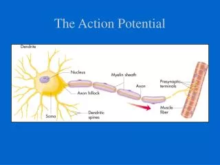
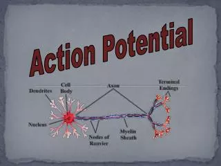
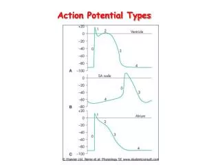
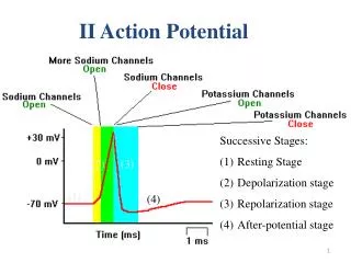
II Action Potential. Successive Stages: Resting Stage Depolarization stage Repolarization stage After-potential stage. (2). (3). (1). (4). Phases of the Action Potential Are Controlled by Gating Mechanisms of Voltage-Gated Ion Channels-

II Action Potential
E N D
Presentation Transcript
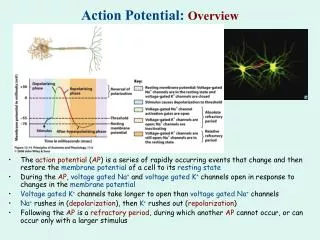
II Action Potential • Successive Stages: • Resting Stage • Depolarization stage • Repolarization stage • After-potential stage (2) (3) (1) (4)
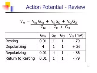
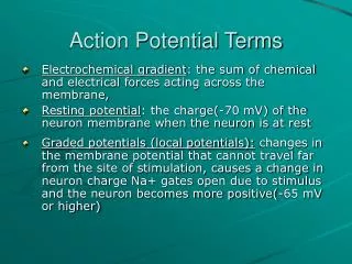
Phases of the Action Potential Are Controlled by Gating Mechanisms of Voltage-Gated Ion Channels- • The depolarizing and repolarizing phases of the action potential can be explained by relative changes in membrane conductance (permeability) to sodium and potassium. • During the rising phase, the nerve cell membrane becomes more permeable to sodium, as a consequence the membrane potential begins to shift more toward the equilibrium potential for sodium which is ? • (ENa = +60mv). • However, before the membrane potential reaches ENa, sodium permeability begins to decrease and potassium permeability increases. This change in membrane conductance again drives the membrane potential toward EK,- • Which is?(EK= -97mV), accounting for repolarization of the membrane
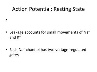
Clinical Focus • -Channelopathies • Voltage-gated channels for sodium, potassium, calcium, and chloride are intimately associated with excitability in neurons and muscle cells and in synaptic transmission. • Channelopathies affecting neurons include episodic and spinocerebellar ataxias, some forms of epilepsy, and familial hemiplegic migraine. • Ataxiasare a disruption in gait mediated by abnormalities in the cerebellum and spinal motor neurons. One specific ataxia associated with an abnormal potassium channel is episodic ataxia with myokymia. In this disease, which is autosomal-dominant, cerebellar neurons have abnormal excitability and motor neurons are chronically hyperexcitable. This hyperexcitability causes indiscriminant firing of motor neurons, observed as the twitching of small groups of muscle fibers, akin to worms crawling under the skin (myokymia). • One of the best-known sets of channelopathies is a group of channel mutations that lead to the Long Q-T (LQT) syndrome in the heart. The QT interval on the electrocardiogram is the time between the beginning of ventricular depolarization and the end of ventricular repolarization. In patients with LQT, the QT interval is abnormally long because of defective membrane repolarization, which can lead to ventricular arrhythmia and sudden death
Action Potential Summary • Reduction in membrane potential (depolarization) to "threshold" level leads to opening of Na+ channels, allowing Na+ to enter the cell • Interior becomes positive • The Na+ channels then close automatically followed by a period of inactivation. • K+ channels open, K+ leaves the cell and the interior again becomes negative. • Process lasts about 1/1000th of a second.
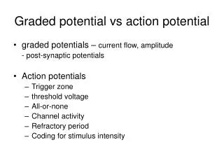
Properties of the Action Potential • “All or none” phenomenon • A threshold or suprathreshold stimulus applied to a single nerve fiber always initiate the same action potential with constant amplitude, time course and propagation velocity. • Propagation • Transmitted in both direction in a nerve fiber
Positive feedback loop graded Na+ potential If YES, then... Reach “threshold”? Stimulation V-gate Na+ channels open Na+ enters (depolarization)
The Speed of Propagation of the Action Potential Depends on Axon Diameter and Myelination After an action potential is generated, it propagates along the axon toward the axon terminal, it is conducted along the axon with no decrement in amplitude. The mode in which action potentials propagate and the speed with which they are conducted along an axon depend on whether the axon is myelinated. The diameter of the axon also influences the speed of action potential conduction: larger-diameter axons have faster action potential conduction velocities than smaller-diameter axons.
Saltatory Conduction • The pattern of conduction in the myelinated nerve fiber from node to node • It is of value for two reasons: • very fast • conserves energy.
MUSCLES-About this Chapter • Skeletal muscle • Mechanics of body movement • Smooth muscle • Cardiac muscle
The Three Types of Muscle Figure 12-1a
The Three Types of Muscle Figure 12-1b
The Three Types of Muscle Figure 12-1c
Skeletal Muscle • Human body contains over 400 skeletal muscles • 40-50% of total body weight • Functions of skeletal muscle-Genrates • Force for locomotion and breathing • Force production for postural support • Heat production during cold stress
Anatomy Summary: Skeletal Muscle Figure 12-3a (2 of 2)
Fascicles: bundles, CT(connective tissue) covering on each one • Muscle fibers: muscle cells
Structure of Skeletal Muscle:Microstructure • Sarcolemma • Transverse (T) tubule • Longitudinal tubule (Sarcoplasmic reticulum, SR) • Myofibrils • Actin(thin filament) • Troponin • Tropomyosin • Myosin(thick filament)
Within the sarcoplasm Triad • Transverse tubules • Sarcoplasmic reticulum -Storage sites for calcium • Terminal cisternae - Storage sites for calcium
Sarcomeres • Sarcomere:bundle of alternating thick and thin filaments • Sarcomeres join end to end to form myofibrils • Thousands per fiber, depending on length of muscle • Alternating thick and thin filaments create appearance of striations
Myosin • Myosin head is hinged • Bends and straightens during contraction
Thick filaments (myosin) • Bundle of myosin proteins shaped like double-headed golf clubs • Myosin heads have two binding sites • Actin binding site forms cross bridge • Nucleotide binding site binds ATP (Myosin ATPase) • Hydrolysis of ATP provides energy to generate power stroke
Thin filaments (actin) • Backbone: two strands of polymerizedglobular actin – fibrous actin • Each actin has myosin binding site • Troponin • Binds Ca2+; regulates muscle contraction • Tropomyosin • Lies in groove of actin helix • Blocks myosin binding • sites in absence of Ca2+
Thick filament: Myosin (head and tail) • Thin filament: Actin, Tropomyosin, Troponin (calcium binding site)
III Molecular Mechanism of Muscular Contraction • The sliding filament model • Muscle shortening is due to movement of the actin filament over the myosin filament • Reduces the distance between Z-lines
Changes in the appearance of a Sarcomere during the Contraction of a Skeletal Muscle Fiber
Energy for Muscle Contraction • ATP is required for muscle contraction • Myosin ATPase breaks down ATP as fiber contracts