Download
1 / 29
290 likes | 940 Vues
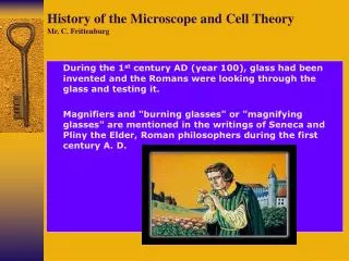
Development of the Light Microscope and the Cell Theory. First compound light microscopes built in mid 1600s by Anton van Leeuwenhoek (Dutch) and Robert Hooke (English); discovered a previously unknown world Corresponded with each other (via letters)

E N D
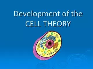
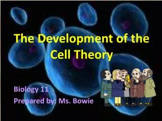
Development of the Light Microscope and the Cell Theory • First compound light microscopes built in mid 1600s by Anton van Leeuwenhoek (Dutch) and Robert Hooke (English); discovered a previously unknown world • Corresponded with each other (via letters) • Robert Hooke coined term “cell” (based on observations of cork) and published the book Micrographia • van Leeuwenhoekfirst to see living, cellular “pond animalcules” • The Cell Theory (mid 1800s; Schleiden, Schwann, and Verchow) 1) All organisms are composed of cells (one or many) • Matthais Schleiden (a botanist) and Theodor Schwann (a zoologist) observed cells in plants and animals, respectively 2) The cell is the basic unit of structure and function in living systems (ex. movement of body results from movement of muscle cells) 3) All cells come from pre-existing cells (Rudolph Verchow) • Spontaneous generation disproved by Francisco Redi and Louis Pasteur; today, oxygenated atmosphere and omnipresence of bacteria result in degradation and loss of free organic materials
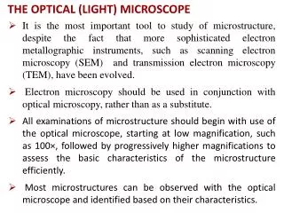
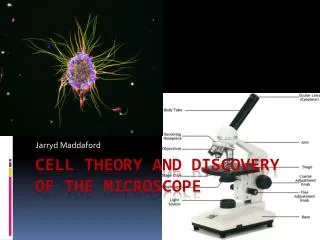
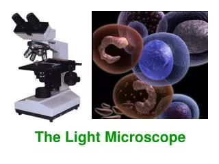
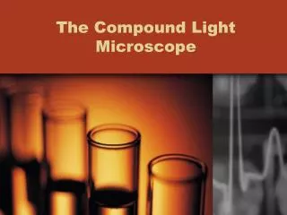
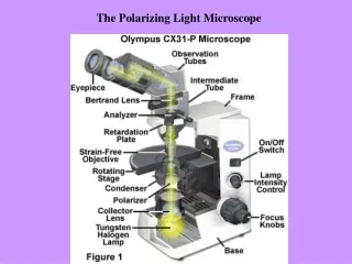
Modern Microscopes • Compound Light Microscope • Magnification a product of the two lenses (eyepiece and objective lens); lowest objective lens known as scanning lens (4X) • Images inverted; resolution limited by relatively large size of photons • Parfocal: object placed in center of field of view prior to changing objective lenses; field of view decreased but object will be there • Dissecting Microscope (Stereomicroscope) • Image is not inverted (better for dissections) • Electron Microscopes (in use by 1950s; incr. resolution) • Scanning Electron Microscope: images are of objects’ surfaces; up to ~60,000x magnifications • Transmission Electron Microscope: images are of sections, internal structures (such as organelles); up to ~200,000x magnifications • Scanning Tunneling Microscopes (Atomic Force Microscopes): images of large molecules possible; multiple technologies; magnifications up to ~100 million x actual size
Cell Types and Shared Structures • Prokaryotic Cells (Prokaryotes: Eubacteria and Archaea) • Most 1-10 μm; seen in fossil record by 3.5 bya; lack a nucleus and other membrane-bound organelles (DNA free in cell, in nucleoid region) • Eukaryotic Cells (Eukaryotes: Fungi, Protists, Plants, and Animals) • Most 10-100 μm; seen in fossil record by 2.2 bya; have a nucleus and other membrane-bound organelles • All Cells Share (thus common ancestor had …) • Cell (plasma) membrane: a boundary; micelles can form naturally • Ribosomes: composed of proteins and RNA; bacterial ribosomes have a different size and structure than those in eukaryotes • DNA, RNA, and the Genetic Code: bacterial chromosome a simple ring of DNA; in eukaryotes, DNA is packaged with proteins • Other molecules / structures: membrane proteins (ex. ATP, ATP synthase), metabolic enzymes, cytoskeletal tubules and filaments
Eukaryotic Structures and Organelles • Structures and Organelles • Nucleus, Nucleolus, and Ribosomes • Nucleus: bound by porous nuclear membrane; contains DNA (chromatin), nucleotides, and nucleolus • Nucleolus: dense, protein-rich area in nucleus; ribosomes form • Ribosomes: in Rough ER and cytoplasm; site of protein assembly (amino acids joined by peptide bonds) • Endoplasmic Reticulum • Golgi Apparatus (Complex) and Vesicles • Lysosomes and Vacuoles • Plastids (chloroplasts, chromoplasts, and mitochondria) • The Cytoskeleton and the Cytosol • Flagella and Cilia
Eukaryotic Structures and Organelles • Structures and Organelles • Nucleus, Nucleolus, and Ribosomes • Endoplasmic Reticulum (ER) • Membranous extension from nuclear membrane; extends throughout cell; transports materials through cell • Rough ER: studded with ribosomes; proteins assembled (esp. membrane and secretory proteins) • Smooth ER: synthesis of lipids (incl. steroids), modification of proteins (incl. detoxification of poisons) • Golgi Apparatus (Complex) and Vesicles • Lysosomes and Vacuoles • Plastids (chloroplasts, chromoplasts, and mitochondria) • The Cytoskeleton and the Cytosol • Flagella and Cilia
Eukaryotic Structures and Organelles • Structures and Organelles • Nucleus, nucleolus, and ribosomes • Endoplasmic Reticulum • Golgi Apparatus (Complex) and Vesicles • Products from ER modified (“tagged”) and transported (“shipped”) via vesicles • In secretory cells, the cell’s main product sent to cell membrane, where vesicles fuse, and products enter blood or saliva • Lysosomes and Vacuoles • Plastids (chloroplasts, chromoplasts, and mitochondria) • The Cytoskeleton and the Cytosol • Flagella and Cilia
Eukaryotic Structures and Organelles • Structures and Organelles • Nucleus, Nucleolus, and Ribosomes • Endoplasmic Reticulum • Golgi Apparatus (Complex) and Vesicles • Lysosomes and Vacuoles • Lysosomes: membrane-enclosed sacs of digestive enzymes found almost exclusively in animal cells • Involved in digestion of food, programmed cell death, immunity, and destruction of cellular waste products • Vacuoles: membranous sacs that bud from the ER, Golgi, or cell membrane • Storage vacuoles store food or water (large central vacuole in plant cells), contractile vacuoles control water balance in some Protists • Plastids (chloroplasts, chromoplasts, and mitochondria) • The Cytoskeleton and the Cytosol • Flagella and Cilia
Eukaryotic Structures and Organelles • Structures and Organelles • Nucleus, Nucleolus, and Ribosomes • Endoplasmic Reticulum • Golgi Apparatus (Complex) and Vesicles • Lysosomes and Vacuoles • Plastids (chloroplast, chromoplast, and mitochondrion) • Chloroplasts: found in many plant and algal cells; contain chlorophyll and perform photosynthesis • Chromoplasts: contain other pigments (ex. melanin in dermis) • Mitochondria: found in all eukaryotes; site of cell respiration (cell’s energy factories) • The Cytoskeleton and the Cytosol • Flagella and Cilia
Eukaryotic Structures and Organelles • Structures and Organelles • Nucleus, Nucleolus, and Ribosomes • Endoplasmic Reticulum • Golgi Apparatus (Complex) and Vesicles • Lysosomes and Vacuoles • Plastids (chloroplasts, chromoplasts, and mitochondria) • The Cytoskeleton and the Cytosol • Cytoskeleton: network of fibers in cell; supports organelles, maintains cell shape, controls movement of some cells; dynamic – can dismantle in one area and be re-assembled; consists of microtubules, intermediate filaments, and microfilaments • Cytosol: the semi-fluid medium of the cell’s cytoplasm • Flagella and Cilia
Eukaryotic Structures and Organelles • Structures and Organelles • Nucleus, Nucleolus, and Ribosomes • Endoplasmic Reticulum • Golgi Apparatus (Complex) and Vesicles • Lysosomes and Vacuoles • Plastids (chloroplasts, chromoplasts, and mitochondria) • The Cytoskeleton and the Cytosol • Flagella and Cilia • Motile appendages in Protists and sperm cells; used for food capture in some Protists; mechanosensory functions in “hair cells” of cochlea and lateral line; clean trachea and bronchi of mucus; found in lining of Fallopian tubes • Ultrastructure includes internal and peripheral microtubules; rotor at base most studied in prokaryotic flagellum • Flagellum: relatively long, often singular; move in undulatory whiplike motion • Cilia: relatively short and found in groups, move in unison
Evolution of the Eukaryotic Cell • Serial Endosymbiotic Theory (Lynn Margulis) • Evolution of new species by the acquisition and incorporation of other organisms’ genomes (a process) • “I picture genes and their products flowing through a sea of cells” (Carl Woese, on early cellular life) • An endosymbiont gradually (over generations) “loses identity” to become an organelle or structure of a larger cell • Aerobic bacterium mitochondrion • Cyanobacterium (photosynthetic) chloroplast • Spirochaete (spirillum bacterium) flagellum • Evidence very strong for origin of plastids • Approximate size of bacteria with structural similarities • Membrane bound, with bacterial proteins in membranes • Contain DNA in ring (bacterial chromosome) • Divide by binary fission • Progressive stages in the “loss of identity” have been observed in various Protists