GIT protozoa
GIT protozoa. Ameba : Entamoeba histolytica Entamoeba dispar Entamoeba coli Entamoeba hartmanni Endolimax nana Iodamoeba bütschlii Flagellates : Giardia lamblia Dientamoeba fragilis Chilomastix mesnili Trichomonas hominis Trichomonas vaginalis (other body sites) Enteromonas hominis
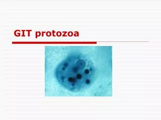

GIT protozoa
E N D
Presentation Transcript
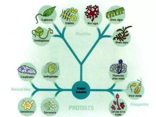
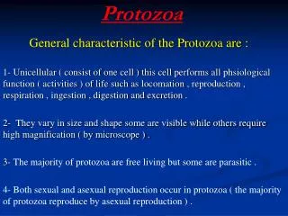
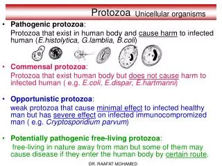
Ameba: • Entamoeba histolytica • Entamoeba dispar • Entamoeba coli • Entamoeba hartmanni • Endolimax nana • Iodamoeba bütschlii • Flagellates: • Giardia lamblia • Dientamoeba fragilis • Chilomastix mesnili • Trichomonas hominis • Trichomonas vaginalis • (other body sites) • Enteromonas hominis • Retortamonas intestinalis • Apicomplexa: • Cryptosporidium hominis • Cryptosporidium parvum • Other: • Blastocystis hominis • Balantidium coli INTESTINAL PROTOZOA unicellular eukaryotic organisms
Fecal-Oral Transmission Factors • poor personal hygiene • food handlers • institutions • children in day care centers • developing countries • highly endemic • poor sanitation • travelers diarrhea • water-borne epidemics • zoonosis • Entamoeba = no • Cryptosporidium = yes • Giardia = controversial • Control/Prevention • improve personal hygiene • especially institutions • treat asymptomatic carriers • eg, family members • health education • hand-washing • sanitation • food handling • protect water supply • treat water if questionable • boiling • iodine • not chlorine
Amoebiasis • Amoebiasis is caused byEntamoeba histolytica. • The organism formerly known asE. histolyticais now known to consist of two distinct species: E. histolytica, which is pathogenic, andE. dispar, which is non-pathogenic. • Cysts of the two species are identical, but can be distinguished by molecular techniques after culture of the trophozoite. E. histolyticacan be distinguished from all amoebae exceptE. dispar, and from other intestinal protozoa, by microscopic appearance. • Amoebiasis occurs world-wide, although much higher incidence rates are found in the tropics and subtropics.
Cont. • The organism exists both as a motile trophozoite and as a cyst that can survive outside the body. • Cysts are transmitted by ingestion of contaminated food or water, or spread directly by person-to-person contact. • Trophozoites emerge from the cysts in the small intestine and then pass on to the colon, where they multiply.
Clinical features • Many individuals can carry the pathogen without obvious evidence of clinical disease (asymptomatic cyst passers). • This is may be due in some cases to the misidentification of non-pathogenic E. dispar as E. histolytica, and it is not clear how often true E. histolytica infection is symptomless. • In affected people E. histolytica trophozoites invade the colonic epithelium, probably with the aid of their own cytotoxins and proteolytic enzymes.
Cont. • The parasites continue to multiply and finally frank ulceration of the mucosa occurs. • If penetration continues, trophozoites may enter the portal vein, via which they reach the liver and cause intrahepatic abscesses. • This invasive form of the disease is serious and may even be fatal.
ulcers with raised borders • little inflammation between lesions
Incubation period • The incubation period of intestinal amoebiasis is highly variable and may be as short as a few days or as long as several months. • The usual course is chronic, with mild intermittent diarrhoea and abdominal discomfort. This may progress to bloody diarrhoea with mucus, and is sometimes accompanied by systemic symptoms such as headache, nausea and anorexia. • Less commonly, infection may present as acute amoebic dysentery, resembling bacillary dysentery or acute ulcerative colitis.
Complications • Complications are unusual, but include toxic dilatation of the colon, chronic infection with stricture formation, severe haemorrhage, amoeboma, and amoebic liver abscess. • Amoebomas, which develop most commonly in the caecum or rectosigmoid region, are sometimes mistaken for carcinoma. They may bleed, cause obstruction or intussuscept. • Amoebic liver abscesses often develop in the absence of a recent episode of colitis. Tender hepatomegaly, a high swinging fever and profound malaise are characteristic, although early in the course of the disease both symptoms and signs may be minimal.
Diagnosis • Microscopic examination of fresh stool or colonic exudate obtained at sigmoidoscopy is the simplest way of diagnosing colonic amoebic infection. • To confirm the diagnosis motile trophozoites containing red blood cells must be identified: the presence of amoebic cysts alone does not imply disease. Sigmoidoscopy and barium enema examination may show colonic ulceration but are rarely diagnostic. • The amoebic fluorescent antibody test is positive in at least 90% of patients with liver abscess and in 60-70% with active colitis. Seropositivity is low in asymptomatic cyst passers.
Management • Metronidazole 800 mg three times daily for 5 days is given in amoebic colitis; a lower dose (400 mg three times daily for 5 days) is usually adequate in liver abscess. • Tinidazole is also effective: dehydroemetine and chloroquine are alternative drugs, but are rarely used. After treatment of the invasive disease, the bowel should be cleared of parasites with a luminal amoebicide such as diloxanide furoate.
prevention • Amoebiasis is difficult to eradicate because of the substantial human reservoir of infection. The only progress will be through improved standards of hygiene and better access to clean water. • Cysts are destroyed by boiling, but chlorine and iodine sterilizing tablets are not always effective.
Thank you Dr. Ayham Abulaila