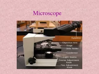
Microscope
Microscope. bla. The samples. Sample 4:. Sample 5:. Sample 7: new wire. Location of wires:. SEM picture. SEM picture. Wire samples: 7 Magnification: Location: Good wire. SEM picture. Wire samples: 4 and 5 Magnification: 40x Location: at beginning of dark region,


Microscope
E N D
Presentation Transcript
Microscope • bla
Sample 4: Sample 5: Sample 7: new wire
SEM picture Wire samples: 7 Magnification: Location: Good wire
SEM picture Wire samples: 4 and 5 Magnification: 40x Location: at beginning of dark region, just below scratch scratch Wire sample 5
SEM picture Wire sample: 5 Magnification: 5000x Voltage: 12 kV Location: at beginning of dark region, just below scratch
SEM picture Wire sample: 5 Magnification: 5000x Voltage: 12 kV Location: 0.5 mm from beginning of dark region
Magnification: x100 (Picture made at the UvA)
Typical EDS spectrum DCSN0014.JPG 12:07 Only peaks of Au and W W: MZ1 1.68 keV Au: Ma 2.12, 2.20 keV
DCSN0026.JPG 12:39 Analysis of dark spot (16kV) DCSN0028.JPG 12:44
DCSN0039.JPG 14:28 Difference dark/light spot • Dark spot: more W • Light spot: more Au 2) 1) We don’t understand peak at 1.40 keV The only element there is Se
Check with WDS for Silicon (Wavelength Dispersion) DCSN0041.JPG 14:38 W
Checked EDS of good wire DCSN0046.JPG 16:16 • 2 places wire sample 7 (new) • 1 place wire sample 6 (non-irradiated) • 1 place wire sample 5 (far from irr) • all identical EDS spectra
Checked EDS of wire sample 5Every 100 um systematically DCSN0048.JPG 16:31 • Normal • A bit of W • A bit of W picture • A bit of W • A tiny bit of W • More W picture • A bit of W • A bit of W • A tiny bit of W • A bit of W DCSN0051.JPG 16:55
Checked for Oxygen with WDS DCSN0054.JPG 17:07 DSCN0057.JPG 17:20
Check with WDS for Carbon: Yes… PS. Also checked for Cr: not found.