Comprehensive Guide to Light Microscopy Equipment
Learn about the types of light microscopes, their components, magnification power, resolution, and aberrations. Discover different types of microscopy, including bright field, fluorescence, dark field, phase contrast, and electron microscopy.

Comprehensive Guide to Light Microscopy Equipment
E N D
Presentation Transcript
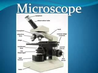
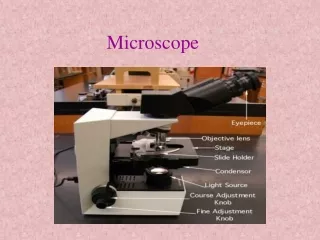
Microscopy • Microscopes are basically classified by the type of light source used: • Bright Field Microscopy: • The light source lies in the visible region. • Basic Components: • Light source (tungsten lamp) • Eyepiece lens: monocular or biocular which has a magnification of 10 X. • Objective lenses: may be four, five or more, 4X, 10X, 40X, and 100X. • The optical tube, which occupies the distance between the eyepiece lens, and the objectives, which is usually equal to 160 mm.
Basic Components • Stage • Sub-stage condenser, which directs the beam of light from the source onto the specimen, it consists of two lenses. • Iris diaphragm, number of leaves can be open or closed to control the amount of light passed. • Microscope contains a built in light source at its base, which usually contains a transformer for adjusting the light intensity.
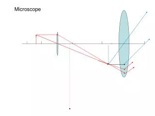
Important points around the light microscope: • Total magnification= • Eyepiece magnification X Objectives magnification. • The image seen by the eye through a compound microscope is termed the virtual image and is upside and reversed. • The numerical aperture (NA): • Is a designation of the amount of light entering the objective from? • The microscopic field • NA = R sin • Where: • R is the refractive index of glass • is the angle made by one ray passing through • the edge of the lens with the other ray passing the center of the lens. • Since sin = AC/AP • Then NA depends on the radius of the lens. Lens A c B Object
Resolving power: Is the useful limit of magnification, it is the ability of microscope, at specific magnification to distinguish two separate objects situated close to one another and the ability of the lens to reveal fine details. The smaller the distance between the two specific objects that can be distinguished apart, the greater the resolution power of the microscope. Minimal distance between two objects = (0.612 X ) / NA The larger NA, the smaller the resolvable distance and hence, the more efficient the resolution power.
Depth of field: Is the capacity of the objective lens to focus in different planes at the same time. This is largely dependent on the NA. Where the greater the NA, the smaller the depth of the field, it is possible to increase the depth of the field slightly by closing the iris diaphragm. Thus decrease the NA. Chromatic Aberration: since light is formed of several wavelength then light component are not bent in the same way as they pass through the lens and therefore are not brought to the same focus.
Spherical aberrations: the light wave, as they travel through the lens, are bent differently, depending in which part of the lens they pass through, rays passing through the peripheral portions of the lens are brought to a shorter focal point than those rays passing through the thicker part of the lens. To correct chromatic and spherical aberration achromatic (brings 2 colors) and apochromatic (brings 3 colors blue, yellow and red) lenses may be used which are fine lenses produced to bring rays of several colors to a common focus
The medium between the objective and the object is a factor that must be taken into consideration for the most effective use of the microscope lens. The low power objective 4x, 10x, and high dry objective 40x use air. When oil immersion lenses are employed, a drop of oil should be used, otherwise bending of the light waves occurs since oil has the same RI of glass, while air increase diffraction.
Types of Microscopes • Ultraviolet Microscopy: • The shorter wavelength of UV can extend the limit of microscope resolution to about 0.1 m. However, UV light is invisible to the human eye, so the image must be recorded on a photographic plate or fluorescent screen. Because this light is absorbed by glass, all lenses must be made of quartz, such microscopes are two expensive for routine use. • Fluorescence microscopy: • A sample labeled with a fluorescent dye is illuminated with UV light, the location of the dye in the specimen is revealed by its fluorescence or emission of visible light
Types of Microscopes • Dark field Microscopy: • One sees a black background; against which suspended bacteria or element appear bright. The dark field microscope uses a special condenser that illuminate the sample with a hallow cone of light in such a manner the light is not directed into the objective lens, revealing the shape of that object. • Phase contrast Microscopy: • Bacterial or animal cells are difficult to be seen using the light microscope unless the sample is dried and stained. This microscope enhances the slight difference in refractive index between the cells and the medium and thus can be used to visualize the living bacteria and platelets, in which the slight differences in RI are converted to differences in light intensity.
Types of Microscopes • Electron Microscopy: • Since magnification greater than 1500X to 2000X are not practical with the light microscope due to decreased efficiency in resolving power. The electron microscope has come into use, where magnification of 50,000X may be obtained, with a high degree of resolving power. • There are two types of electron microscope: • Transmission Electron Microscope (TEM) {2 dimensional} • Scanning Electron Microscope (SEM) {3 dimensional}