Download
1 / 69
690 likes | 714 Vues
Learn about how the invention of the microscope revolutionized science and how it led to the development of the Cell Theory. Discover the different types of microscopes and their capabilities, as well as the structures and functions of cells. Explore the differences between prokaryotic and eukaryotic cells, and understand the importance of the cell membrane.

E N D
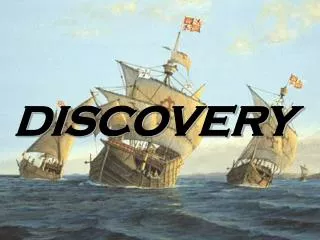
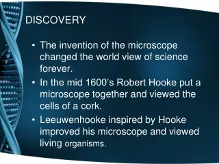
DISCOVERY • The invention of the microscope changed the world view of science forever. • In the mid 1600’s Robert Hooke put a microscope together and viewed the cells of a cork. • Leeuwenhooke inspired by Hooke improved his microscope and viewed living organisms.
microscopes • Light microscope - light passes through one or more lenses to produce an enlarged image of a specimen. Can magnify up to 2,000X. Can view living organisms. • Stains and fluorescent dyes can be added to show specific structures in the cell. • Optical techniques can show image in 3D and computer processing can follow movements of cell parts and materials in and out of the cell. • Image is sharp when 0.2 µm
Your compound Light microscope • Ocular lens 10X • Low Power 4X multiply 10X ocular Total magnification is 40X • Medium is 10X multiply 10 X Total magnification is 100X • High Power 40X multiply 10X Total magnification is 400X
Microscope • Magnification increases the size of an object. • As the magnification increases your field of view decreases. • Resolution increases the ability to see details of the speci.men
Electron microscope- Forms an image of a specimen using a beam of electrons rather than light. Can magnify up to 200,000X. Must be done in a vacuum. Living organisms can not survive in a vacuum. TEM-Transmission electron microscope shines a beam of electrons through a thin specimen. SEM-Scanning tunneling microscope runs a pencil like beam of electrons back and forth across the surface of the specimen which produce dramatic three dimensional images.
SEM Sickle Cell Virus
SEM Images Dust Mite Pollen
Cell Theory • Hooke and Leeuwenhooke recorded all that they saw. • Schleiden, Swann, & Virchow further studied cells and proposed The Cell Theory. Page 55.
Cell Theory • All living organisms are composed of one or more cells. • Cells are the basic structure and organization of all living organisms. 3. Cells come from previously existing cells, with cells passing copies of their genetic material on to their daughter cells.
Cell Size • A cells size is limited by the ratio of their outer surface and their inner volume. • As cells increase their volume (the stuff inside) Their out membrane stretches (like blowing up a balloon). • Too much volume • Can burst the cell. • Reduce transport across the outer membrane.
Cell Shape • Cells can vary in shape depending on their function • All cells contain organelles which are similar in function to human internal organs. • The cell membrane surrounds and protects the cell.
Two Types of Cells • Eukaryotic • Membrane bound nucleus that houses all of the cells genetic material • Many membrane bound organelles (p.58). • Prokaryotic • No Membrane bound nucleus - the cells genetic material floats randomly in the cell • Many organelles NOT bound by a membrane • Have a cell wall surrounding the cell membrane
Cell membrane Cytoplasm Cell membrane Cytoplasm Internal Organization Prokaryotic Cell Eukaryotic Cell Nucleus Organelles
Eukaryotes Prokaryotes Nucleus Endoplasmic reticulum Golgi apparatus Lysosomes Vacuoles Mitochondria Cytoskeleton Cell membrane Contain DNA Ribosome Cytoplasm Compare and Contrast
Prokaryotic Examples ONLY Bacteria
Prokaryotes • Cell wall in a bacterial cell is typical made from a carbohydrate-protein complex called peptoglycan or similar substance. • Cytoplasm and cytoskeleton • Do not contain a nucleus DNA is circular and DNA area is called nucleoid • Some bacteria contain plasmids DNA that are independent of the main DNA. Bacteria can exchange these. Ex. Plasmid may express allow a bacteria to express an ability like make a sugar outer coating. • Have ribosomes • Cilia and flagella for movement.
Venn Diagrams Animal Cells Plant Cells Cell Wall Chloroplasts Cell membrane Ribosomes Nucleus Endoplasmic reticulum Golgi apparatus Lysosomes Vacuoles Mitochondria Cytoskeleton Centrioles Compare and Contrast
Organelles • Cell membrane • Nucleus • Mitochondrion • Ribosome • Endoplasmic reticulum • Golgi apparatus • Lysosome/peroxisomes • Microfilament & microtubules • Cilia and flagella • Cell wall – Plant cell • Vacuole – plant cells • Chloroplast – plant cells
Check Point 1.What is the importance of a surface area to volume ratio? 2. Compare the structure of a eukaryotic cell with a prokaryotic cell? 3.When Hooke first used the word cell, did the intend to have it apply to living material? Explain. 4. Name two structures that all cells have.
Cell membrane Fluid Mosaic ModelProposed in 1972 by Singer • Selectively permeable & pliable membrane • Membrane lipids • Phospholipids – polar head and non-polar tails. The polar head are hydrophilic and the non-polar tails are hydrophobic • Steroid (cholesterol) is located between the tails of the phospholipids
Cell Membrane • Proteins embedded in cell membrane • Peripheral – external peripheral proteins have a carbohydrate attached • Integral – in between the phospholipids; can form channels to allow transport in and out of the cell
Cell Membrane • Boundary of the cell • Made of a phospholipidbilayer
Nucleus • Is surrounded by the nuclear matrix which is a stiff membrane • There is an inner membrane called a nuclear envelope • Chromatin (a combination of DNA and protein) is inside the nucleus. The chromatin coils and becomes chromosomes when the cell is ready to divide.
Nucleus • Nucleus stores genetic information • Is where RNA is copied from DNA • Nuclear pores allow RNA to travel out of the nucleus. • Contains the nucleolus where ribosome's are made.
Nucleus • Control center of the cell • Contains DNA • Surrounded by a double membrane • Usually the easiest organelle to see under a microscope • Usually one per cell
Ribosome • Most abundant in the cell • No outer membrane • Is made up of a protein and RNA • Are made in the nucleus • Can be free floating (remain in cell)or attached to endoplasmic reticulum (for export). • Make proteins • Found in all cells
Ribosome • Site of protein synthesis • Found attached to rough ER or floating free in cytosol • Produced in a part of the nucleus called the nucleolus
Endoplasmic reticulum (ER) • Highway of tubes and sacs by which molecules move around the cell • Has a membrane • Two types • Rough – is covered in ribosomes is found in cells that make many proteins • Smooth – helps make steroids, regulates calcium in muscles, and breaks down toxins in the liver
Endoplasmic Reticulum • A.k.a. “ER” • Connected to nuclear membrane • Highway of the cell • Rough ER: studded with ribosomes; it makes proteins • Smooth ER: no ribosomes; it makes lipids
Golgi Apparatus • Processing, packaging and secretion center • Is a system of tubes and sacs • Enzymes that are in the Golgi attach carbohydrates and lipids to the proteins. • Make cell membrane components or the cell. • Packages for distribution lysosomes.
Golgi Apparatus • Looks like a stack of plates • Stores, modifies and packages proteins • Molecules transported to and from the Golgi by means of vesicles
Lysosomes • Spherical organelles that contain hydrolytic enzymes which digest proteins, nucleic acids, lipids and carbohydrates. • This allows lysosomes to recycle cell parts. • Lysosmoes remain in the cell.
Lysosomes • Garbage disposal of the cell • Contain digestive enzymes that break down wastes Which organelles do lysosomes work with?
lysosomes • Pump enough H ion to maintain a pH of 5. • Contain about 40 different enzymes that breakdown macromolecules.
Peroxisomes • Globular organelles found in almost all eukaryotes but many different types place where oxidation reactions take place. • ex. Breaks down Hydrogen peroxide. • ex. Brake down of ethanol • ex. Breakdown of fatty acids
Mitochondria • Change organic compounds to energy called adenosine triphosphate (ATP) • Has two membranes • Inner membrane called cristae which increases the surface area so more compounds can be converted to ATP. • Outer membrane protects and allows transport. • Has its own DNA and can reproduce to make more mitochondria
Mitochondria • “Powerhouse of the cell” • Cellular respiration occurs here to release energy for the cell to use • Bound by a double membrane • Has its own strand of DNA (circular)
Cilia and Flagella • Both organelles are on the exterior of the cell • Assist with movement • Cilia – many short hair-like structures that beat in unison to cause movement • Flagellum – one long tail-like structure that whips to cause movement