Download
1 / 85
880 likes | 1.17k Vues
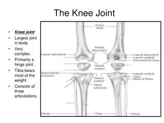
The Knee: Anatomy and Injuries. 2 Joints at the Knee. Tibiofemoral Joint – formed between the femur (femoral condyles), and the tibial plateau Patellofemoral joint – formed between the patella and the femur. Bones of the Knee. Femur proximal – head and neck of femur, greater trochanter

E N D
2 Joints at the Knee Tibiofemoral Joint – formed between the femur (femoral condyles), and the tibial plateau Patellofemoral joint – formed between the patella and the femur
Bones of the Knee Femur proximal – head and neck of femur, greater trochanter distal – medial and lateral condyles and epicondyles
Bones of the Knee Patella – largest sesamoid bone in body Tibia – tibial plateau forms knee joint with femur The fibula is not a part of the knee joint
Quadriceps tendon: connects Quads to patella Patellar Ligament: connects patella to tibia
The Quadriceps: Muscles of Knee Extension 1. Vastus Medialis 2. Vastus Lateralis 3. Vastus Intermedius 4. Rectus Femoris – 2 joint muscle that also acts as a hip flexor
The Hamstrings Muscles of Knee Flexion: 1. Biceps Femoris 2. Semimembranosus 3. Semitendinosus
The Adductors (Groin) • Muscles that Adduct the thigh 1. Adductor Longus 2. Adductor Magnus 3. Adductor Brevis 4. Gracilis
Sartorius Muscle Flexes, abducts, and laterally rotates thigh Longest muscle in the body, “tailor’s muscle” Crosses hip and knee joint
The Iliotibial Tract (IT Band) - neither a muscle or tendon, but a long, thick band of tissue that inserts into the lateral tibia (Gerdy’s Tubercle)
The Major Knee Ligaments ACL – Anterior Cruciate Ligament PCL – Posterior Cruciate Ligament MCL – Medial Collateral Ligament LCL – Lateral Collateral Ligament
The Cruciate LigamentsThe major stabilizing ligaments of the knee ACL Runs from posterior femur to anterior tibia Prevents anterior displacement of tibia PCL Runs from anterior femur to posterior tibia Prevents posterior displacement of tibia
The Collateral Ligaments MCL: Medial Collateral Ligament Runs from medial femur to medial tibia Prevents valgus force LCL: Lateral Collateral Ligament Runs from lateral femur to head of fibula Prevents varus force
The Meniscus A “c”-shaped piece of fibrocartilage located in the knee joint between the femur and attached to the top of the tibia Cartilage = meniscus
Differences between medial and lateral Medial - larger and more C-shaped - more firmly attached to tibia - has attachments to MCL Lateral - smaller and more round or O-shaped - not firmly attached to tibia and LCL
Blood Supply to the Meniscus Mostly avascular – little or no blood supply Only the outer 20% has a blood supply * Does not have the ability to heal itself unless there is a small tear in the outer 20%
Functions of Meniscus Stability Shock absorption Lubrication and nutrition Allows adequate weight distribution
Normal Torn
Good Knee Anatomy Review Website http://academic.pgcc.edu/~aimholtz/AandP/PracPrac/Muscle/ThighLeg/ThighLeg.html
Total Knee Joint Replacement Surgery to replace a painful damaged or diseased knee joint with an artificial joint (prosthesis) Artificial hip invented 1962 1969 – first artificial knee in USA
The Knee Surgery Thin layer of bone removed from femur – thin metal replaces it Upper layer of tibia replaced with plastic Back of patella replaced with plastic Parts fastened with “bone cement”
Risks of Knee Joint Replacement Blood clots in large veins Infection Stiffness Implant Loosening/Failure - more of a problem in younger patients
Genu Valgum: “knock knees”
Genu Varum: “bowlegs”
Genu Recurvatum: hyperextension of the knee joint
Patellar Tracking Disorder When your patella is out of balance or the patellar cartilage is damaged, you can have knee pain while climbing stairs, running, standing up from a bent-knee position, squatting, or even sitting for a period of time. This kind of pain, called anterior knee pain or patellofemoral pain syndrome, is sometimes caused by a common kneecap problem known as patellar tracking disorder.
Causes Quadriceps weakness Tendon and muscle tightness in the leg, foot, or hip areas Improper athletic technique or training A blow to the kneecap Excessive body weight, which overstresses the knee joint. Genetics
Genetics (cont.) - An excessively long patellar tendon - patellar shape, hip structure, or a shallow femoral groove for the patella to glide along
Patellofemoral Disorders Problems with patella – most common cause of knee pain Anatomy: - Patella is a sesamoid bone formed in Quad tendon - Patellofemoral joint – patella and femur - Compression forces – <body weight during walking 2.5 x body weight during stairs
Patellar Tendonitis “Jumper’s Knee” Inflammation and degeneration of the tendon that connects the kneecap (Patella) to the shin bone (Tibia).