Download
1 / 45
1.09k likes | 3.84k Vues
Please click audio icon to hear Carol’s narration. Mycology Dermatophytes. Division of Medical Technology Carol Larson MSEd, MT(ASCP). Click icon for audio. Basic Characteristics. Medium growth rate (1-3 weeks) Identification Colony morphology Microscopic morphology

E N D
Please click audio icon to hear Carol’s narration MycologyDermatophytes Division of Medical Technology Carol Larson MSEd, MT(ASCP)
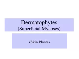
Click icon for audio Basic Characteristics • Medium growth rate (1-3 weeks) • Identification • Colony morphology • Microscopic morphology • Hyphae – hyaline & septate • Macroconidia, Microconidia • Physiological tests • Clinical significance – Tinea (ringworm)
Click icon for audio Clinical Significance • Skin • Tinea corporis • Tinea pedis • Tinea cruris
Click icon for audio Clinical Significance • Hair • Tinea capitis • Tinea barbae • Ectothrix • Endothrix
Click icon for audio Clinical Significance • Nails • Tinea unguium
Click icon for audio Clinical Significance
Click icon for audio Epidemiology • Anthropophilic • Man • Zoophilic • Animals • Geophilic • Soil
What three body sites do the dermatophytes infect? Hair, skin and nails
What is the difference between endothrix and ectothrix? Endothrix means the mold has conidia inside the hair shaft, whereas Ectothrix means the conidia are only on the outside of the hair shaft.
What infection do the dermatophytes cause? Tinea (also referred to as “ringworm”). Another term that can be used is dermatophytosis.
Click icon for audio Laboratory Diagnosis • Specimen collection • Direct examination • Culture • Identification
Click icon for audio Laboratory Diagnosis Specimen Collection • Hair • Plucked, not cut, from edge of lesion • Skin • Wash, scrape from margin of lesion • Nails • Scrapings from nail bed or infected area • Transport in sterile petri dish
Click icon for audio Laboratory Diagnosis Direct Examination • Examine hair for fluorescence • Wood’s lamp • Yellow green fluorescence = positive
Click icon for audio Laboratory Diagnosis Direct Examination • Examine specimen for fungal elements • 10% KOH preparation • Calcofluor white stain
Click icon for audio Laboratory Diagnosis Specimen processing • Hair • Cut into short segments • Nails • Mince into small pieces
Click icon for audio Laboratory Diagnosis Culture Media • Select two media types • General purpose – Sabouraud’s agar • Selective – Mycosel agar • Antibiotics • Gentamicin: inhibits normal bacterial flora • Cycloheximide: inhibits saprophytic fungi
Click icon for audio Laboratory Diagnosis Culture Growth Requirements • Place specimen pieces on culture media • Can streak for isolation • Incubate at 30°C in ambient (room) air • Growth at 3 days to 3 weeks • Examine plates frequently for 4 weeks
Click icon for audio Laboratory Diagnosis Identification • Colony morphology • Microscopic morphology • Scotch tape preparation • Tease prep • Slide culture
Click icon for audio Laboratory Diagnosis Identification • Physiologic tests • Urea hydrolysis • Hair perforation • Rice grain media • Vitamin requirements
How can hair, skin and nails be evaluated directly for fungal elements? Wood’s lamp fluorescence (hair only), 10% KOH preparation, and Calcofluor white fluorescent stain.
What are the incubation requirements when suspecting a dermatophyte infection? Fungal media is incubated at 30°C in ambient air for 4 weeks. There is one exception and that is Trichophyton verrucosum that requires 35ºC.
What primary procedures are performed to identify the dermatophytes? Colony morphology, microscopic morphology (Scotch tape prep, tease prep, or slide culture), and physiologic tests such as urea hydrolysis and hair perforation.
Click icon for audio Etiologic Agents • Microsporum species • Epidermophyton species • Trichophyton species
Click icon for audio Microsporum canis • Colony morphology:
Click icon for audio Microsporum canis • Microscopic morphology:
Click icon for audio Microsporum gypseum • Colony morphology:
Click icon for audio Microsporum gypseum • Microscopic morphology:
Click icon for audio Microsporum audouinii • Colony morphology:
Click icon for audio Microsporum audouinii • Microscopic morphology:
How can Microsporum species be differentiated from each other microscopically? Characteristic appearance of the macroconidia, and the general appearance of the hyphae (such as pectinate bodies). As a group, Microsporum have few to absent microconidia.
Click icon for audio Epidermophyton floccosum • Colony morphology:
Click icon for audio Epidermophyton floccosum • Microscopic morphology:
Click icon for audio Trichophyton rubrum • Colony morphology:
Click icon for audio Trichophyton rubrum • Microscopic morphology:
Click icon for audio Trichophyton rubrum • Physiological tests • Urea: negative • Hair perforation: negative
Click icon for audio Trichophyton mentagrophytes • Colony morphology: Downy Granular Velvet
Click icon for audio Trichophyton mentagrophytes • Microscopic morphology:
Click icon for audio Trichophyton mentagrophytes • Physiologic tests: • Urea: positive • Hair perforation: positive
How can Trichophyton mentagrophytes be differentiated from Trichophyton rubrum? Urea hydrolysis and hair perforation tests. T. mentagrophytes is positive for both, and T. rubrum is negative for both.
How can Microsporum, Epidermophyton, and Trichophyton species be differentiated microscopically? Microsporum has numerous thick-walled macroconidia with RARE microconidia, Epidermophyton has numerous club-shaped macroconidia hanging out in groups of 2-3 with NO microconidia, and Trichophyton has thin-walled macroconidia and MANY microconidia.
Click icon for audio Dermatophytes In Summary … • Causes Tinea (ringworm) • Medium growth rate = 1-3 weeks • Grows on Mycosel agar • Identification • Colony morphology, microscopic exam, and physiologic tests • Etiologic agents • Microsporum, Epidermophyton, Trichophyton species
Who am I? Potato Dextrose Agar Reverse LPCB Stain of Slide Culture Microsporum canis
Who am I? Potato Dextrose Agar LPCB Stain of Slide Culture Epidermophyton floccosum
Who am I? Hair Perforation Potato Dextrose Agar LPCB Stain of Slide Culture Trichophyton mentagrophytes
Who am I? Potato Dextrose Agar Reverse LPCB Stain of Slide Culture Microsporum gypseum