Download
1 / 68
690 likes | 936 Vues
Understand femur fractures, their symptoms, statistics, and risks, including diagnosis and treatment options for effective care. Learn about the anatomy of the femur and the importance of early intervention in cases of fractures.

E N D
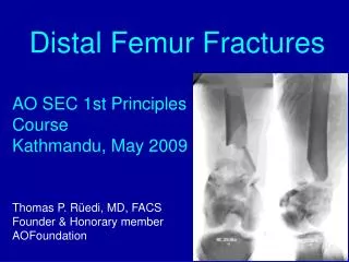
The Fractured Femur January/ February 2013 issue of Radiologic Technology Directed Readings In the Classroom
Instructions: • This presentation provides a framework for educators and students to use Directed Reading content published in Radiologic Technology. This information should be modified to: • Meet the educational level of the audience. • Highlight the points in an instructor’s discussion or presentation. • The images are provided to enhance the learning experience and should not be reproduced for other purposes.
Introduction • The femur is the largest and strongest bone in the human body, and great force is necessary to fracture it. Radiography is the gold standard for diagnostic imaging of femurs, but diagnosis can be complicated with nondisplaced or occult fractures. Particularly in the emergency setting, modalities such as magnetic resonance imaging or computed tomography may be necessary. Effective treatment of femur fractures is needed to restore homeostatic function and prevent complications.
Symptoms • In the case of high-impact trauma, it is often obvious that the femur has been fractured. Stress or insufficiency fractures that are not pronounced may be apparent by pain in the upper leg or hip, and it may be extremely painful or even impossible for an individual to place any weight on the affected leg. The fractured leg may be deformed or shorter than the opposite leg, and the femur may even rip through the skin on the thigh in cases of severe fracture.
Symptoms • Fractures of the hip, including the femoral head or femoral neck, are often evidenced by pain in the hip, knee, or lower back. Other common, and somewhat more specific, symptoms include the inability to stand or walk and the foot on the affected side turning at an abnormal angle, making the leg look shorter than the leg on the unaffected side of the body.
Statistics and Risks • As many as 250,000 hip-joint fractures occur in the United States each year, and the majority of hip fractures are the result of falling. More than one-third of Americans older than 65 years of age fall each year, and 90% of all hip fractures occur in individuals older than age 50. Approximately 15% to 25% of elderly individuals who suffer a hip fracture die within 1 year.
Statistics and Risks • Up to 90% of femoral shaft fractures in adolescents and the general population are caused by motor vehicle accidents, including cars, bicycles, or being stuck by a vehicle as a pedestrian.Femur fractures also often result from sporting accidents; illness or disease that affects bone integrity, such as vitamin D deficiency, systemic lupus erythematosus, or cancer; and certain medications, particularly long-term use of bisphosphonates for osteoporosis and cancer-related metastases.
Statistics and Risks • Femoral neck stress fractures occur most often in highly active individuals, such as elite distance runners, military recruits, and dancers. Another group with a disproportionately high risk of femoral neck stress fractures includes postmenopausal women and individuals with conditions resulting in loss of bone mineral density, including osteopenia, osteoporosis, Paget disease, and hyperparathyroidism. Stress fractures to the femoral neck are uncommon in the general public and exceedingly rare in children.
Statistics and Risks • Although most femur and hip-joint fractures in the general population are the result of an accident, certain factors can increase the risk of these fractures in the elderly population, including: • Low bone mineral density. • Bone-related medical conditions, such as osteoporosis or cancer-related bone metastasis. • Conditions that make an individual more prone to falling, such as dementia and visual impairment. • Personal and familial (maternal) history of fracture.
Statistics and Risks • Excessive consumption of alcohol or caffeine. • Physical inactivity. • Low body weight. • Tall stature. • Medications that affect bone mineral density, such as psychotropicsand long-term use of bisphosphonates. • Similar to the pediatric population, femur fracture in the elderly can be a sign of abuse, and any suspicion of abuse should be reported to the proper authorities.
Statistics and Risks • More than 250 000 subcapital hip fractures below the femoral head are reported annually in the United States, costing an estimated $15 billion per year.By 80 years of age, an estimated 20% of white women and 10% of white men will suffer a hip fracture, most of which are secondary to osteoporosis.The number of individuals older than 65 years is estimated to rise from approximately 35 million in 2000 to nearly 71 million by 2030, and with the aging population it is estimated that the incidence of femur and hip fractures in the elderly will increase from 12.4% in 2000 to 19.6% by 2030.
Anatomy - Skeletal • The femur is the upper bone of the leg. Its function is to allow for walking by connecting the hip to the knee. The femoral head is a ball that fits into the socket joint of the hip at the acetabulum. This ball-and-socket system is held in place by ligaments, or ligamentumteresfemoris. The femoral neck connects to the shaft of the femur at an angle to allow for ambulation. The femur is almost completely cylindrical. Like other long bones, the femur consists of a body and 2 extremities (upper and lower). The upper (proximal) extremity is made up of the femoral head, neck, and greater and lesser trochanters. The lower (distal) extremity is cuboid and consists of 2 oblong projections called the lateral condyle and the medial condyle. The condyles are separated by the recessed patellar surface that connects with the bones of the knee.
Anatomy - Skeletal • The femoral head fits into the hip socket and is round. It is directed upward, medialward, and slightly forward. There is a depression on the femoral head, called the fovea capitisfemoris, where the head attaches to the ligamentumteres.The weight of the human trunk rests on the 2 femoral heads. The femoral neck connects the femoral head to the femoral body. The neck is a flattened pyramidal process of bone that forms an angle opening medialward. This angle is widest in infancy and becomes narrower as an individual ages. In adults, the femoral neck forms a 125° angle with the body.The structure of the femoral head and neck allows transmission of body weight, as well as the load-bearing and torsion associated with walking.
Figure 1. Illustration of the anterior (A) and posterior (B) femur. • A
Types of Fractures • A stress fracture is an overuse injury that results when the muscle becomes too fatigued to absorb shock and transfers the overload of stress to the underlying bone, and may not be evident on initial radiographs. On radiographs, it often appears as a sclerotic band across the bone, although a defined fracture line is invisible. In such cases, radionuclide bone scanning is possibly the best imaging modality; stress fractures appear as areas of increased uptake before any changes are visible on radiographs. Similarly, pathologic fractures occur spontaneously in abnormal bone, particularly in the presence of bone tumors. Often, a bone lesion is obvious; sometimes the borders of the lesion can be poorly defined, and diagnosis requires recognizing the irregularity of the margins of the fracture. Imaging other parts of the skeleton may help to confirm or rule out suspicion of bone metastases.
Types of Fractures • Fatigue fractures result from abnormal stress being placed on normal bone.Compression fractures occur when bone collapses either because of excessive pressure or illness. These types of fractures occur most commonly in the vertebrae, sacrum, pubic rami, and femoral neck. Salter-Harris fractures are those that occur to the growth plates, almost always due to force. Nonaccidental fractures are linked with abuse, particularly in children but also in the elderly. Radiologic technologists, and interpreting physicians should be aware of potential abuse, which is often indicated by multiple fractures, particularly fractures that appear to have taken place at different times. Other factors pointing to nonaccidental injury include epiphyseal and metaphyseal fractures, which often appear on radiographs as small chips from the long bones (corner fractures) that likely result from twisting or pulling the limbs.
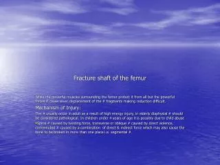
Classifying Fractures • Clinicians have created several systems to classify fractures of the femur, femoral head, femoral neck, and growth plates. Fractures can be located on the proximal, middle, or distal third of the femoral shaft,and they can be characterized in several ways, according to: • The direction of the fracture line. • The relationship of the bone fragments involved. • The number of fragments involved. • Any exposure to the outside air.
Classifying Fractures • The direction of the fracture line often depends on the type of force causing the trauma. Transverse fractures are horizontal breaks across the femoral shaft and are caused by a force exerted perpendicular to the shaft. Force applied in the same direction as the long axis of the bone produces diagonal or oblique fractures across the shaft. Longitudinal fractures also occur along the long axis of the bone. Finally, spiral fractures encircle the femoral shaft and are produced by a torque injury.
Classifying Fractures • The fracture can display 1 or a combination of the variations in position: • Displacement – misalignment of the fragments; represents the distance the distal fracture fragment is offset from the proximal fracture fragment. • Angulation – the amount of deviation from the normal angle of the distal and proximal fragments. • Shortening – the amount of overlap in the ends of the fracture fragments. • Rotation – the extent the fracture fragments pivot or turn from normal position.
Classifying Fractures • Salter-Harris fractures involve the femoral growth plates, either alone or combined with an adjacent part of the femur. This classification system includes 5 types and is particularly important because of its predictive value. Type I and type II fractures are associated with a good prognosis, whereas type IV and type V can result in early fusion of the epiphysis and subsequent shortening of the bone.
Classifying Fractures • Fractures of the hip are classified according to 4 subtypes: intertrochanteric fractures, subtrochanteric fractures, femoral head fractures, and femoral neck fractures. An intertrochanteric fracture is the most common type of hip fracture and involves a fracture line between the greater and lesser trochanters. Intertrochanteric fractures also carry the best prognosis of any hip fracture, particular in healthy individuals (see Figure 3). Subtrochanteric fractures occur at the femoral shaft below the lesser trochanter.
Classifying Fractures • Femoral neck fractures are categorized according to the Garden Classification, which includes: • Type 1 – stable; involves a minor crack in the femoral neck. • Type 2 – involves a complete crack in the femoral neck but no bone displacement. • Type 3 – a displaced fracture with the fragments remaining connected to one another; also may involve rotation of the bone fragments, angulation, or both. • Type 4 – completely displaced with no connection between the fractured fragments; likely to disrupt blood supply to the femoral head.
Classifying Fractures • The PipkinClassification is the most widely used classification criteria for femoral head fractures (see Box 2). According to this system, fractures are categorized into 4 types based on increasing severity. The classifications influence treatment decisions and predict outcomes. For example, type I fractures typically have better outcomes and often are managed without surgery, using physical therapy and limited weight bearing. The occurrence and severity of complications increase from type I to type IV. Salter-Harris fractures involve the femoral growth plates, either alone or combined with an adjacent part of the femur. This classification system includes 5 types and is important because of its predictive value. Type I and type II fractures are associated with a good prognosis, whereas type IV and type V can result in early fusion of the epiphysis and subsequent shortening of the bone.
Diagnostic Imaging - Radiography • With bone fracture, radiographs remain the gold standard for diagnostic imaging. In the United States, nearly every patient who presents to the emergency department with a suspected fracture undergoes plain-film radiography.Not only can radiographs be used to visualize fracture or dislocation, they also can help determine whether underlying bone is normal or whether a fracture occurred because of an abnormality (ie, a pathological fracture). Radiography can be used to distinguish a fracture from other conditions or diseases, such as cancer and bone metastases, osteomyelitis, Paget disease, or dislocation. Radiography also can show the position of the bone ends before and after treatment of a fracture, and it can be used to assess healing and complications.
Diagnostic Imaging Radiography • With bone fracture, radiographs remain the gold standard for diagnostic imaging. In the United States, nearly every patient who presents to the emergency department with a suspected fracture undergoes plain-film radiography.Not only can radiographs be used to visualize fracture or dislocation, they also can help determine whether underlying bone is normal or whether a fracture occurred because of an abnormality (ie, a pathological fracture). Radiography can be used to distinguish a fracture from other conditions or diseases, such as cancer and bone metastases, osteomyelitis, Paget disease, or dislocation. Radiography also can show the position of the bone ends before and after treatment of a fracture, and it can be used to assess healing and complications.
Diagnostic Imaging - Radiography • In the case of bone trauma, at least 2 projections are required: anteroposterior (AP) and lateral. Sometimes a fracture or dislocation will be seen on only 1 radiograph and would be missed without the second image, especially when fracture fragments are minimally displaced. The position of a fracture must be assessed with more than 1 image. For the AP projection, the patient should be positioned supine with the femur centered over the film or image receptor and a table top or Bucky. The central ray should be midshaft. For the lateral projections of the distal femur, the technologist would use a cross-table lateral projection. The opposite leg should be pulled up and over the affected leg, centered with the film or image receptor and a table top or Bucky. The central ray should be midshaft. If the image does not include both joints, another radiograph should be taken of the other joint.
Diagnostic Imaging - Radiography • Even with 2 projections, fractures may be invisible on radiographs, in which case additional projections should be performed at the discretion of the radiologist as follows: • Oblique projections. • Stress images, which are taken with a joint under stress to show that it is unstable, as in the case of dislocation. • Flexion and extension projections, which should only be taken with the individual performing the movement and not on an unconscious individual. • Radiographs of the nonfractured side for comparison.
Diagnostic Imaging - Radiography • Intracapsular fractures (ie, those above the trochanters) usually are apparent on radiographs; however, when a fracture is not evident and clinical suspicion of a fracture is high, magnetic resonance (MR) imaging may be preferred.
Diagnostic Imaging- Radiography • Often, acute injuries to the growth plates are not clearly visible because of the cartilaginous-osseous composition and irregular contours of the physes.The epiphyseal growth plate appears on radiographs as a white boundary, making it easily confused with an impacted fracture in which trabeculae have become enmeshed. Because radiographs may show physeal widening as the only sign of displacement, imaging of the unaffected leg may help with diagnosis via comparison, particularly with type I Salter-Harris fractures. In the case of type V Salter-Harris fractures, changes to the bone often are missed on plain radiographs.Although varus and valgus stress projections may be indicated to demonstrate separation between the epiphysis and the metaphysis, particularly in injuries around the knee, stress maneuvers may cause further damage in other individuals and should be performed with caution.
Diagnostic Imaging - Radiography • Femoral neck fractures often occur in elderly individuals because of falls. In the elderly population in particular, it is important to distinguish between fractures of the pelvis and nondisplaced, impacted, or occult fractures of the femoral neck. Radiographic imaging of the femoral neck should include AP images and images of the lateral ipsilateral femur with internal rotation.Because a patient should not be turned on their side if they have a fractured femoral neck, a cross-table lateral view is needed. A fracture of the femoral neck appears on radiography as dark streaks across the bone where the continuity of cortical and spongy bone is interrupted.
Diagnostic Imaging - Radiography • Radiographic imaging of the hip should include an AP and a lateral projection. The AP image should be captured with the patient supine and the foot internally rotated 15°to secure the best view of the femoral neck. The central beam should be directed toward the femoral head. The x-ray tube should be positioned 100 cm from the focal plane of the image receptor to produce an image at 20% magnification. The cross-table lateral projection should be taken when an individual is suspected of having a hip fracture or dislocation. For the cross-table lateral projection, the patient should be supine, with the opposite hip flexed and abducted. The cassette should be placed against the lateral side of the affected hip, and the central beam directed horizontally toward the groin, with approximately a 20°angle of cephalic tilt.
Diagnostic Imaging - Magnetic Resonance Imaging • MR imaging plays an important role in the diagnosis of femur fractures. MR can be used to assess soft-tissue damage resulting from femur fracture, and is superior to CT in its ability to visualize soft tissue. MR imaging also is used to assess tissue composition and image lesions in multiple planes and to accurately delineate geographic relationships of the body’s internal structures. In the emergency setting, MR remains the imaging modality of choice for occult hip and pelvic fractures. Hip fractures often are missed on radiography.MR imaging is indicated in nonambulatory patients with negative radiographic images, and both insufficiency fractures and stress fractures appear as characteristic bone marrow edema on MR imaging. Detecting hip fractures early ensures patients are properly treated and minimizes complications that result from prolonged immobilization and hip surgery.
Diagnostic Imaging - Magnetic Resonance Imaging • In a case report and literature review by Cheon et al, the utility of MR imaging in diagnosing insufficiency fractures of the femur not immediately evident on radiography was evaluated. Radiographic images were compared with MR images, and the results indicated that 4 of the 7 individuals studied had impending fractures, as indicated on MR imaging. The presence of the femoral cortical ridge on radiography appeared as a complete transverse fracture line on MR imaging, leading the authors to postulate that this finding indicated the potential for fracture and possible need for prophylactic fixation. Moreover, the authors stated that although MR imaging is the most effective modality for diagnosing stress fractures of the tibia and femur in athletes, it is also effective in evidencing impending fractures in women at risk for such injury as a result of long-term bisphosphonate use.
Diagnostic Imaging - Magnetic Resonance Imaging • In a case report and literature review by Cheon et al, the utility of MR imaging in diagnosing insufficiency fractures of the femur not immediately evident on radiography was evaluated. Radiographic images were compared with MR images, and the results indicated that 4 of the 7 individuals studied had impending fractures, as indicated on MR imaging (see Table 1). The presence of the femoral cortical ridge on radiography appeared as a complete transverse fracture line on MR imaging, leading the authors to postulate that this finding indicated the potential for fracture and possible need for prophylactic fixation. Moreover, the authors stated that although MR imaging is the most effective modality for diagnosing stress fractures of the tibia and femur in athletes, it is also effective in evidencing impending fractures in women at risk for such injury as a result of long-term bisphosphonate use.
Diagnostic Imaging - Magnetic Resonance Imaging • MR imaging is not as effective as radiography or CT in showing fracture lines because cortical bone does not produce an MR signal. However, MR imaging is use ful in showing bony injuries often not obvious on CT scans, such as bone bruises that do not involve cortical bone disruption but do result in hemorrhage and edema in the bone marrow. This is because bone bruises, for example, cause hemorrhage within the bone that replaces the marrow with fat, thereby altering the MR signal.An altered signal may be visible in the presence of a hemorrhage, and a bone bruise may be visible even without a discernible fracture on a radiograph.
Diagnostic Imaging - Magnetic Resonance Imaging • MR imaging can depict altered arrest lines and transphyseal bridging abnormalities before they are evident on radiographs. In addition, coronal MR imaging can be used to image a fracture to the femoral neck not visible on radiographs of the hip.With fractures of the growth plate, MR imaging is the most accurate modality in showing fracture anatomy in the acute phase of injury (ie, the first 10 days following injury).On T1-weighted MR imaging, fractures appear as a transverse band of low-intensity (bright signal) marrow replacement.On T2-weighted imaging, fractures appear as high signal surrounding edema.
Diagnostic Imaging - Computed Tomography • Compared with radiography, CT has the distinct advantage of more clearly showing obvious fracture lines as well as cortical abnormalities associated with nondisplaced fractures. CT can show fracture lines in greater detail, as well as the position and orientation of fracture fragments, better than other imaging modalities. CT particularly is useful for imaging fractures in complex skeletal structures, such as the hip, face, shoulder, and foot. CT can produce sagittal and coronal reformations, as well as 3-D models of the injured area, that display the position and orientation of major fracture fragments and allow for viewing of the bone as if the soft tissues were removed.Although CT has been shown to be less sensitive than MR imaging in detecting fractures, CT is recommended for individuals who cannot tolerate MR imaging.
Diagnostic Imaging - Computed Tomography • With fractures around the hip, CT can show the relationship of the fragments to the joint as well as any loose fragments in the joint. CT also demonstrates tissue damage and hematomas that can result from fractures. CT is often quicker and more comfortable, requiring less body manipulation than radiography, making it advantageous for seriously injured individuals, such as those involved in a car accident. Melvin et al conducted a study to assess the utility of CT for detecting and managing femoral neck fractures. Researchers found that the addition of CT, as well as modification of the Garden Classification as nondisplacedvs displaced, improved intraobserver reliability in interpreting the findings in the 5 cases included in the study. Classification of femoral neck fractures was more likely to change when CT was combined with radiography, and agreement regarding classification was improved with the addition of CT.
Diagnostic Imaging - Nuclear Medicine • Radionuclide bone scanning (also referred to as bone scintigraphy) with technetium 99m-labeled diphosphonate tracer material plays a role in diagnosing fractures because of its high sensitivity compared with other imaging modalities. The diphosphonates accumulate rapidly in the bone, and almost 50% of the tracer is absorbed by the skeletal system within 2 to 6 hours after injection. The rate of uptake of the radiotracer depends on blood flow and new bone formation. Thus, radionuclide bone scanning can be used to show signs of bone healing, as well as cell turnover and other physiologic signs of fracture.
Diagnostic Imaging - Nuclear Medicine • Radionuclide bone scanning also is used to diagnose bone tumors or cancer, rule out a bone infection or avascular necrosis, and evaluate metabolic disorders that affect the bones (eg, osteoporosis or Paget disease), thereby distinguishing such conditions from a fracture.For example, on bone scans, osteomyelitis almost always appears as a combination of focal hyperfusion, focal hyperemia, and focally increased bone uptake.Because this modality can be used to show cell turnover, it is useful for examining skeletal lesions.Bone metastases typically appear on bone scans as multiple foci of increased activity, although they occasionally appear as areas of increased uptake of tracer material.
Diagnostic Imaging -Nuclear Medicine • Positron emission tomography using 18F-fluorodeoxyglucose (FDG-PET) scanning can be used to examine an individual for abnormal processes in the bone; its use in detecting femur fractures is limited. FDG-PET measures metabolic activity and molecular function via injected contrast material that is absorbed into the body, emitting radiation that is detected by the PET scanner. Because cancerous cells grow more rapidly than normal cells, the high absorption of the glucose-based contrast material by malignant cells allows for cancer diagnosis. The reported sensitivity of FDG-PET in detecting bone metastases is approximately 90%. In the case of femoral fracture, FDG-PET may be helpful in distinguishing fracture from bone metastases. FDG-PET has been shown to have a high false-positive rate, is more expensive than standard radionuclide bone scanning, and less readily available than MR imaging.
Diagnostic Imaging - Nuclear Medicine • Single photon emission computed tomography (SPECT) is another type of nuclear medicine examination that uses radiotracer material to generate gamma decay to obtain images. Like CT, its images can be formatted in multiple planes. With femur fracture examination, SPECT may assist in evaluating femoral neck stress fractures in conjunction with planar scintigraphy.In a retrospective study of 38 individuals in the military, 33 had undergone planar scintigraphy with SPECT before MR imaging of the hip for the evaluation of femoral neck fracture. When SPECT was added to planar scintigraphy, sensitivity rose from 50% to 92.3% (P = .03), and accuracy in detecting high-grade fractures improved from 12.5% to 70% (P = .025). When SPECT is an option, the authors suggested that it be added to bone scintigraphy for evaluating individuals with suspected femoral neck fractures.
Dual-energy X-ray Absorptiometry • Dual-energy x-ray absorptiometry (DXA) may have a potential role in detecting femur fractures, although it most commonly is used to detect changes in bone density. Bazzocchi et al retrospectively reviewed 739 DXA examinations to detect collateral findings and understand their effect on patient care. The review included 231 examinations of the lumbar spine, 221 of the femur, 191 of the whole body, and 96 of the vertebrae. Incidental findings were noted in 117 of the DXA examinations, 27 of which involved the femur. Of these incidental findings, 50 were confirmed using another imaging technique. None of the incidental findings were mentioned in the DXA reports. Based on these results, the authors stressed that when DXA is used to evaluate bone integrity or possible fracture, interpreting clinicians should evaluate DXA images for their potential in qualifying collateral findings related to diagnosis.
Diagnostic Imaging • PET scan coupled with CT (PET-CT) has shown promise as a potential imaging modality for individuals with osteoporosis and atypical femoral shaft fractures because of its usefulness in defining certain aspects of pathogenesis, site specificity, and potential prodromal abnormalities, in addition to providing insight about radiokinetic variables of skeletal blood flow and markers for bone formation. However, more studies on the clinical use of PET-CT as a diagnostic imaging modality for femur fracture are warranted.
Diagnostic Imaging • Ultrasonography plays a limited role in bone assessment, mostly in the imaging of joint effusions, blood flow, and the presence of foreign bodies within the soft tissues.Ultrasound-guided femoral nerve blocks may be used in the emergency setting to achieve adequate analgesia for severe femoral fractures. • Finally, high-resolution peripheral quantitative computed tomography (HR-pQCT) has shown promise in recent years because of its ability to image bone density and isotropic voxel size, which may prove helpful in assessing osteoporosis and fracture risk as well as treatment efficacy.