Download
1 / 8
500 likes | 2k Vues
Histology of Adrenal Gland - Medulla. outer cortex (the main part of the adrenal glands) inner medulla (which accounts for about 10% of the adrenal glands) surrounded by a thick connective tissue capsule

E N D
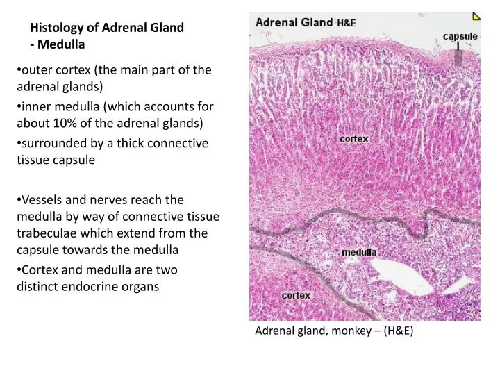
Histology of Adrenal Gland - Medulla outer cortex (the main part of the adrenal glands) inner medulla (which accounts for about 10% of the adrenal glands) surrounded by a thick connective tissue capsule Vessels and nerves reach the medulla by way of connective tissue trabeculae which extend from the capsule towards the medulla Cortex and medulla are two distinct endocrine organs Adrenal gland, monkey – (H&E)
Adrenal gland, medulla (H&E) boundary between cortex and medulla (arrowheads) is sharp and distinct because of the staining quality of the cells on both sides nothing separates the cells of the cortex (zonareticularis) from cells of the medulla
Adrenal gland, medulla (H&E) consists of anastomosing cords and trabecuale of groups of cells (arrow) separated by venous sinusoids (arrow with tail) medullary cells a.k.apheochromocytesor chromaffin cells, synthesize and store catecholamines epinephrine and norepinephrine- released into the venous sinusoids upon signals by sympathetic neurons whose axons form synapses with pheochromocytes venous sinusoids are lined by a fenestrated endothelial layer.
Adrenal gland, medulla (H&E) groups of pheochromocytes are bounded by supporting cells (sustentacular cells) and reticular fiber network The sustentacular cells are flat to spindle-shaped and not readily identifiable with routine H&E staining The pheochromocytes form part of the neuroendocrine system and can be regarded as postganglionic sympathetic neurons.
Adrenal gland, medulla (H&E) Granules in pheochromocytes (arrow) range in size, with smaller ones containing epinephrine and the larger ones norepinephrine Chromogranin proteins package catecholamines in granules.
Adrenal gland, medulla (H&E) medulla contains single or groups of sympathetic ganglions (arrows) and preganglionic sympathetic nerve bundles (arrowheads) coming from splanchnic nerves The preganglionic nerve axons synapse with pheochromocytes (arrow with tail and squiggly arrow).
Adrenal gland, medulla (H&E) Medulla is shown to contain not only pheochromocytes (arrow with tail) but also ganglion cells (arrows on the right) and nerve bundles (arrowhead on the left).
Adrenal gland, medulla (H&E) The sympathetic ganglions are large plump cells with abundant amphophilic cytoplasm (arrowhead) and a nucleus with a prominent nucleolus (thin arrow).