Download
1 / 37
1.82k likes | 6.51k Vues
GINGIVAL RETRACTION. DR. ARAVIND R KUDVA. Definition. Gingival tissue displacement is the deflection of the marginal gingiva away from a tooth.

E N D
GINGIVAL RETRACTION DR. ARAVIND R KUDVA
Definition • Gingival tissue displacement is the deflection of the marginal gingiva away from a tooth. • It is defined as the procedure of temporary eversion (widening of gingival sulcus) or resection of gingiva away from the tooth surface or deepening of gingival sulcus to expose the cervical portion of tooth in order to have proper marginal finish to the restoration or by establishing a good cervical cavosurface margin to the tooth preparation or by recording the preparation accurately.
Indications: • Presence of Sub gingival Caries. • Cervical abrasion or erosion • Elastic impression procedure • Cast restoration • To control haemorrhage and gingival seepage • Esthetic consideration
Subgingival tooth fracture • Subgingival finish lines a) When crown root ratio decreases b) Subgingival caries c) Coronal fracture • Gingival polyps
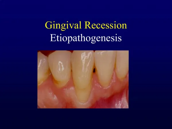
Contraindications: 1. Poor oral hygiene 2. Presence of periodontal disease 3. Gingival recession 4. Periodontal bone loss 5. Gingival disease
Basic Techniques for Gingival Retraction • Mechanical • Chemico mechanical • Surgical: • Electrosurgery • Rotary curettage • Lasers - 980-nm Diode and 1064-nm Nd:YAG
Mechanical Method • Rubber dam b. Cotton twills with ZnoE cement c. Copper Band impressions
Rubber dam • Charbeneauand Gilmore • Heavy weight rubber dams were used. • Produced retraction by compression . Advantages control of seepage and hemorrhage. ease of application. Disadvantages full arch models cannot be made. severe cervical extension preparations.
Cotton twills with ZnoE cement • Employs gentle pressure over a period of time. • ZnoE mixed into creamy consistency, • Cotton twills rolled into this mass and then on a towel to gain compactness. • Prevents sticking of pack to the instruments and gives ease in handling. • Should reflect the tissue laterally. • Pack held in place with fast setting Znoe cement.
Copper band/tube impression A- Oversized copper band should be at least 2.0mm wider than the mesiodistal width of the tooth. B – Band trimmed and contoured inward to allow the band to just clear the preparation margin during impression procedure.
The band is filled with soft wax and seated on the tooth. • The wax is chilled and impression is removed. • The impression indicates over extension of the band. • Adjustments if required may be made and second trial impression is made.
The wax is melted and modelling compound is introduced. • Incisal or occlusal end gingival end • Seat the band securely into its position. • Pressure is applied on the compound directly. • Chill the impression. • A towel clamp may be used to remove the impression.
Strings or fibers of different types, wet or dry: • Plain cotton thread • Unwaxed floss • Cotton cord • 2.0 untreated surgical silk
Chemicomechanical Method • Combines chemical action with pressure packing. • Cords or strings impregnated by chemicals • Various drugs used for gingival displacement include: • 0.1 – 8% - Epinephrine • 100% Alum solution • 5% and 25% aluminium chloride • Ferric subsulfate (Monsel’s solution) • 13.3% Ferric sulfate solution • 8% and 40% zinc chloride solution • 20% and 100% Tannic acid solution
Classification of retraction cords a. Depending on the configuration Twisted Knitted Plain b. Depending on surface finish waxed unwaxed
c. Depending on the chemical treatment Plain Impregnated d. Depending on number strands Single Double-string
e. Depending on the thickness (color coded) black 000 yellow 00 purple 0 blue 1 green 2 red 3
Purpose of Gingival Retraction • To record the unprepared tooth structure immediately adjacent to the margins. • Control of moisture and to obtain a dry field of operation. • To displace the gingiva apically and laterally. • To record the subgingival finish line
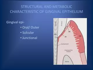
Management and preservation of Biologic width Biologic Width: A band of soft tissue attachment between the base of the gingival sulcus and alveolar crest that is composed of approximately 1mm of junctional epithelium and 1mm of connective tissue.
Referred as Bermuda Triangle of Dentistry. • Violation of biologic width leads to chronic gingival inflammation, pocket formation and osseous defects. • Tooth preparation must terminate at least 2mm coronal to the alveolar crest at the base of gingival sulcus within the intracrevicular space.
Procedure • Isolate the operating area. • Retraction cord of approximately 5 cm (2 inches) is cut with sterile cotton pliers. • Twisted cord – Grasp between thumb and forefinger of each hand and twist to produce tightly wound cord of small diameter. For braided cord- twisting not necessary. • Moisten the cord with aluminum chloride solution.
Form the cord into A “u” and loop it around the prepared tooth. • Gently slip the cord between the tooth and gingiva in mesialinterproximal area with Fischer packing instrument or a De plastic instrument.
Once the cord has been tucked in on the mesial, use the instrument to lightly secure it in the distal interproximal area. • Proceed lingually by starting from the mesiodistal lingual corner around to distolingual corner. • The tip of instrument should be inclined slightly towards the area where the cord has already been placed i.e., mesially.
Instrument must be angled slightly towards the root to facilitate subgingival placement of cord. • Cut off the length of cord protruding from the mesial sulcus as closely as possible to the interdental papilla. • Continue packing the cord around the facial surface, overlapping the cord in mesial interproximal area.
Pack all but the last 2.0 or 3.0 mm of cord. • Place a large bulk of gauge in the patient’s mouth. • After 10 minutes, remove the cord slowly to avoid bleeding.
Rotary Curettage • Definition: It is a technique which, by the use of high speed diamond instrument, facilitates the placement and recording of the subgingival margins of cast restorations.
Suitability of gingiva for use of this method is determined by three factors: • Absence of bleeding upon probing. • Sulcus depth less than 3mm • Presence of adequate keratinised gingiva.
Technique • Sulcus is probed to have an idea of attachment levels and also to examine bleeding on probing. • Using a high speed, Torpedo shaped diamond of 150-180 Grit finish line is extended apically, one half to two-third the depth of sulcus converting the finish line to a chamfer. • Water spray is essential to minimize Trauma. • Cord impregnated with aluminium chloride or alum is gently placed to control Heamorrhage.
Electrosurgery • Definition: Harri’s defines it as the use of specially designed electronic equipment that produces a limited variety of high frequency wave forms for the purposes of cutting or removing soft tissue.
Mechanism of action • Oringer states that heat generated by the resistance of soft tissue to the passage of high frequency electrical energy causes molecular disintegration of cells in the line of surgical incision. • It uses radio currents in the range of 1.5 to 7.5 million cycles/sec.
Important facts • Important facts to be taken into consideration: • Contraindicated in patients with pacemaker or delayed healing because of debilitating disease or radiation therapy. • Not suitable on thin attached gingiva. • Should not be used with metal instruments.
III Recent Advances • 1. Magic foam • 2. Expasyl • 3. Strips