Download
1 / 15
150 likes | 345 Vues
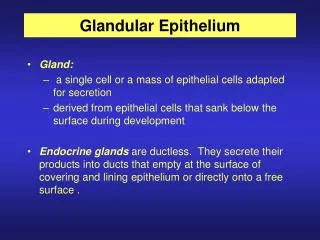
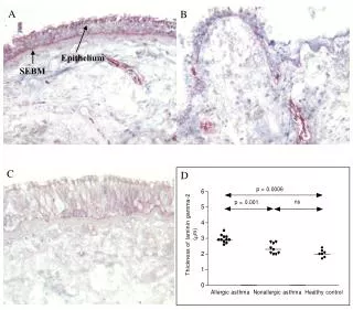
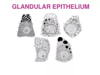
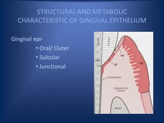
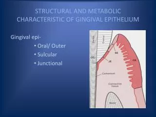
STRUCTURAL AND METABOLIC CHARACTERISTIC OF GINGIVAL EPITHELIUM. Gingival epi- Oral/ Outer Sulcular Junctional. OUTER OR ORAL EPITHELIUM. - Covers the crest and outer surface of the marginal gingiva and the surface of attached gingiva. - Thickness - 0.2-0.3 mm

E N D
STRUCTURAL AND METABOLIC CHARACTERISTIC OF GINGIVAL EPITHELIUM Gingival epi- Oral/ Outer Sulcular Junctional
OUTER OR ORAL EPITHELIUM - Covers the crest and outer surface of the marginal gingiva and the surface of attached gingiva. - Thickness - 0.2-0.3 mm - Keratinized or parakeratinized. -Keratinization diminishes with age & onset of menopause
Order of keratinization- palate > gingiva > ventral aspect of tongue > cheek
Keratins K1, K2 & K10-K12 which are specific to epidermal differentiation, seen more in orthokeratinized than parakeratinized. K6 & K16 characteristic of highly proliferative epithelia,K5 & K14, stratification-specific cytokeratins,are also present. K19 present in parakeratinized,absent in orthokeratized.
SULCULAR EPITHELIUM Lines the gingival sulcus Non keratinized SSE without rete pegs Extends from coronal limit of junctional epithelium to the crest of gingival margin. Shows hydropic degeneration.
Contain – K4, K13 (Oesophageal type cytokeratin) - K19 No Merkel cell Glu-6-phosphate dehydrogenase activity is less. Acid phosphatase is totally absent Lower degree of activity in sulcular epi than in outer epi particularly in the case of enzymes related to keratinization.
Sulcular epithelium has the potential to keratinize if- If it is reflected and exposed to oral cavity. The bacterial flora is totally eliminated.
Sulcular epithelium is extremely imp - it may act as a semipermeable memb via which injurious bacterial products passing to the gingiva & tissue fluid from the gingiva seeps into the sulcus.
Unlike the junctional epithelium - sulcular epithelium is not heavily infiltrated with PMNs. - Less permeable
Junctional Epithelium Consists collar like band of SSE. Nonkeratinized 3-4 layers thick in early life, 10-20 layers thick with increase in age. Tapers from its coronal end. Length 0.25-1.35mm. Express K19, K5, K14
The cells can be grouped in to two strata- JE is formed by the confluence of oral epithelium and the reduced enamel epithelium during tooth eruption. The JE is attached to the tooth surface (epithelial attachment) by means of internal basal lamina. It is attached to gingival connective tissue by an external lamina.
The attachment of the JE to the tooth is reinforced by the gingival fibers which brace the marginal gingiva against the tooth surface, so the JE and gingival fibers are considered a functional unit K/A DENTOGINGIVAL UNIT. Dentogingival unit = JE + Gingival fib
FUNCTIONS OF JE JE is firmly attached to the tooth surface, forming an epithelial barrier against plaque bacteria. It allows access of gingival fluid , inflammatory cells and components of immunological host defense to the gingival margin. exhibit rapid turnover, which contributes to the host-parasite equilibrium and rapid repair of damaged tissue. cells of JE have endocytic capacity equal to that of macrophage and neutrophils and that this activity might be protective in nature.
Cuticular structure on the tooth Cuticle – Acquired coating – (exogenous origin) Developmental - (part of tooth)