GLANDULAR EPITHELIUM
230 likes | 826 Vues
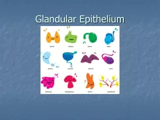
GLANDULAR EPITHELIUM. CLASSIFICATION OF GLANDS. EPITHELIAL SPECIALISATION FOR SECRETION A PATCH OF EPITHELIAL CELLS (INTERNAL SURFACE) OR A DOWNGROWTH THAT PROLIFERATES (EXTERNAL SURFACE) MICROSCOPIC, PART OF AN ORGAN OR AN ORGAN BY ITSELF CONNECTIVE TISSUE FRAMEWORK.

GLANDULAR EPITHELIUM
E N D
Presentation Transcript
CLASSIFICATION OF GLANDS • EPITHELIAL SPECIALISATION FOR SECRETION • A PATCH OF EPITHELIAL CELLS (INTERNAL SURFACE) ORA DOWNGROWTH THAT PROLIFERATES (EXTERNAL SURFACE) • MICROSCOPIC, PART OF AN ORGAN OR • AN ORGAN BY ITSELF • CONNECTIVE TISSUE FRAMEWORK
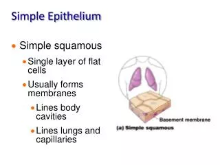
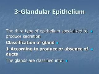
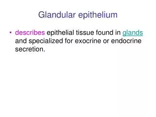
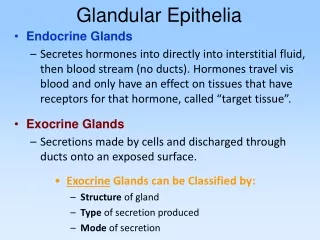
GLANDULAR EPITHELIA • Endocrine glands • Release hormones into surrounding fluid (blood flow) Exocrine glands Secrete through ducts ontothe surface of the gland
MORPHOLOGICAL TYPES • MULTICELLULAR • Organs containing glandularepithelium • (large glands, crypts) • UNICELLULAR • Individual secretory cells • (goblet cells) TUBULO-ACINAR TUBULO-ALVEOLAR TUBULAR ALVEOLAR SIMPLE SIMPLE BANCHING COMPOUND
CLASSIFICATION ACCORDING TO SECRETIONAL TYPES • Merocrine (product released through exocytosis) Cells of the gland are unaltered • Ex: Eccrine sweat glands, salivary glands • Apocrine (involves the loss of both product and cytoplasm) • Ex: Mammary glands, prostate, scent glands • Holocrine (destroys the cell) • Ex: Sebaceous glands
SEROUS SECRETORY CELLS TYPICAL ACINUS (UNIT) TUBULOALVEOLAR small lumenround nuclei wellstained cytoplasm THE CYTOPLASM IS POLARIZED BASAL CYTOPLASM – basophilic due to protein synthetic organelles (ribosomes on rough endoplasmic reticulum APICAL CYTOPLASM - variously-staining secretory vesicles (zymogen granules) The Golgi apparatus is usually located midway along the cell, typically in a supranuclear position.
SEROUS SECRETORY CELLS TYPICAL ACINUS (UNIT) COILED TUBULAR wide lumenround nuclei wellstained cytoplasm THE CYTOPLASM IS POLARIZED TYPICAL SEROUS ACINUS!!! Long excretory portion Opens onto the external surface
MUCOUSSECRETORY CELLS TYPICAL ACINUS (UNIT) wide lumen flat nuclei poorly stained cytoplasm THE CYTOPLASM IS POLARIZED MUCOUS CELLS - "empty" appearance BASAL CYTOPLASM – nucleus and rest of cell organelles MIDDLE and APICAL CYTOPLASM – large secretory granules Mucus does not stain well with standard acidic or basic dyes, but is demonstrated with the Periodic Acid Shiff procedure (PAS stain).
SEROUS AND MUCOUS ACINI THE SEROUS / MUCOUS DISTINCTION IS BASED ON THE CELL'S PRODUCT SEROUS, LIKE SERUM - CLEAR, WATERY SOLUTION OF ENZYMES MUCOUS, LIKE MUCUS - GLYCOPROTEIN MIXTURE MIXED glands (most salivary glands) contain both types of cells (many serous demilunes) SEROUS glands (e.g., parotid gland or pancreas) MUCOUS glands (e.g., Brunner's glands).
UNICELLULAR MUCOUS GLAND GOBLET CELLS : scattered among enterocytes within the epithelium of the small intestine and colon, respiratory and reproductive tracts. They secrete MUCUS, which facilitates passage of material. The name "goblet" refers to the cell's shape, narrow at the base and bulging apically. The apical end of each goblet cell is occupied by a large mass of mucus, which compresses adjacent cells and displaces the nucleus toward the basal end of the cell. As in other mucous cells, the nucleus is compact and intensely-stained. Goblet cells comprise an integral part of the epithelium, attached by junctional complexes (evidenced in light microscopy as the "terminal bar") to adjacent absorptive cells. The proportion of goblet cells to absorptive cells increases along the entire length of the bowel, with relatively few in the duodenum and very many in the colon.
APOCRINE SECRETION 1. APOCRINE SWEAT GLANDS open into hair follicles Large glands FOUND: axillae, perineum, and areolae mammae (phylogenetic remnant of the mammalian sexual scent gland; the sweat is initially odorless upon production. It is only after skin's bacteria has acted upon the apocrine sweat that it develops an odor)
APOCRINE SECRETION 2. TYPICAL ACINUS (UNIT) wide lumen oval nuclei light staining in cytoplasm MAMMARY GLAND NON-LACTATING THE CYTOPLASM IS POLARIZED APOCRINE CELLS - irregular height (torn luminal surface) BASAL CYTOPLASM – nucleus + cell organelles MIDDLE and APICAL CYTOPLASM – large secretory granules (extreme exocytotic procedure- the upper part maybe missing) PROSTATE PSEUDO-APOCRINE LACTATING
HOLOCRINE SECRETION TYPICAL ACINUS (UNIT) no lumen round nuclei light / no staining in cytoplasm • THE CYTOPLASM IS NOT POLARIZED • HOLOCRINE CELLS - gradually age and die • round cells with lipid-filled vacuoles • branched acini join at short duct • duct empties into hair follicle • Associated with hair follicles in hair covered areas • Independent of hair follicles at mucosal margins • (lips, eyelids, penis, labia minora, nipples) • Secretes oily substance (sebum) onto hair • to waterproof hair and skin
STRUCTURES AIDING SECRETION MYOEPITHELIAL MUSCLE BUNDLES MYOEPITHELIAL CELLS ACTIN STAINING