Download
1 / 46
470 likes | 726 Vues
TYPES OF EPITHELIA, GLANDULAR EPITHELIUM. Dr. Katalin Gallatz. FUNCTION AND TYPES OF EPITHELIUM. COVERING EPITHELIUM. 1. Covering and protecting surfaces 2. Diffusion 3. Absorbtion 4. Excretion 5. Secretio n 6. Sensation 7. Contractility. GLANDULAR EPITHELIUM

E N D
TYPES OF EPITHELIA, GLANDULAR EPITHELIUM Dr. Katalin Gallatz
FUNCTION AND TYPES OF EPITHELIUM COVERING EPITHELIUM • 1. Covering and protecting surfaces • 2. Diffusion • 3. Absorbtion • 4. Excretion • 5. Secretion 6. Sensation 7. Contractility GLANDULAR EPITHELIUM SENSORY EPITHELIUM MYOEPITHEL
Covering and protection – Covering epithelium 1. epithelial cells cover the external and internal surfaces, 2. protect underlying tissue from mechanical injury, harmful chemicals, invading bacteria and from excessive loss of water. Diffusion Simple squamous epithelium promotes the diffusion of gases, liquids and nutrients( walls of capillaries and lungs).
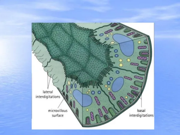
Characteristics of covering epithelia 1. Cells of the epithelium are closely packed. 2. Junctional complexes can be found beetween the cells. 3. Small amount of intercellular substance 4. Cells form one or more layers. 5. Cover or line all internal and external body surfaces. 6. Basement membrane separates it from the underlying tissues
I. Basal lamina • a. lamina lucida • b. lamina densa • II. Reticular lamina BASEMENT MEMBRANE COMPONENTS:
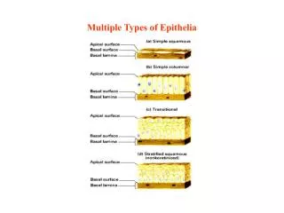
Types of covering epithelium Covering epithelium can be divided into two groups depending on the number of layers 1. simple epithelium is only one cell thick 2. stratified epithelium is two or more cells thick
Simple epithelia: simple squamous simple cuboidal simple columnar pseudostratified ciliated columnar simplesquamous simplecuboidal pseudostratifiedcolumnar simplecolumnar
Simplesquamousepithelium and simplecuboidalepitheliumfromkidney
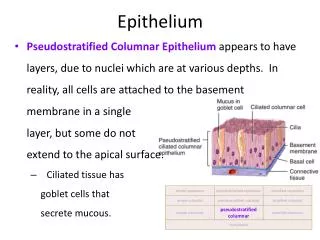
simplecolumnar and pseudostratifiedcolumnarepithelium columnar cells basal cell
Stratified epithelia stratified squamous non keratinised stratified squamous keratinised stratified cuboidal stratified columnar transitional epitheliu
Stratifiedsquamousepithelium non keratinized keratinized
Stratified columnar epithelium 3. Stratum columnare 2. Stratum spinosum 1. Stratum basale
Transitional epithelium (urothelium) 3.Umbrella cell layer 2. Pear- shaped cells 1. Stratum basale
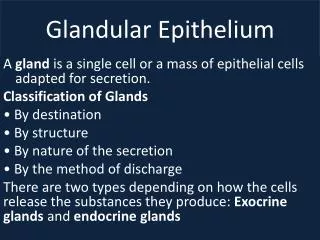
Glandular Epithelium • Epithelialare cells specialized to produce secretion.. • Secretion – exocytotic release of products, (not metabolic wastes) • Molecules to be secreted may be stored in membrane bound secretory granules
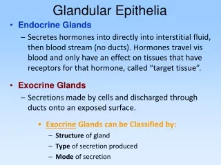
Glands are classified as : • ENDOCRINEandEXOCRINE depending on their route of secretion
ENDOCRINE GLANDS • Secretory products called hormones, are secreted directly into the blood • No ducts
Exocrine glands can be classified according to the A) Number of cells Uni- or multicellular B) Mode of secretion holocrine, apocrine, merocrine D) Shape tubular, alveolar (acinar), tubuloalveolar C) Secretory products mucous and serous
EXOCRINE GLANDS • release their products onto the free surface of the skin or mucous membranes • mucouos membranesof tubular organsof the digestive, respiratory or reproductive tracts.
Classification accordingto the number of the cells 1.Unicellular 2. Multicellular A.) simple (one unbranched duct)- sweat gland B.) compound (branched ducts) parotid gland
Unicellular glands • They are scattered among other non-secretory epithelial cells • They have no ducts, but they secrete their products directly on the free surface • The most common unicellular exocrine glands are the goblet cells (mucus secreting cells)
Unicellular endoepithelial gland:goblet cell Narrow basal part: nucleus and RER basophilic Apical part: mucin containing secretory granules foamy apperence From Dr. Zita Puskár
Goblet cells trachea small intestine simple columnar epithelium with cuticule Pseudostratified columnar epithelium From Dr. Zita Puskár
Demonstration of mucins with differentstainings small intestine PAS large intestine mucicarmin small intestine H.E. redish pale purple From Dr. Zita Puskár
Multicellular glands a) intraepithelial gland gland is entirely within the epithelium. b) extraepithelial gland below epithelium
Multicellular endoepithelial glands In the epithelium of the male urethra From Dr. Zita Puskár
Compound tubuloalveolar mixed gland submandibular gland
Modes of Secretion(how products leave the cell) • merocrine - secretion does not affect the well-being of the cell sweat glands, salivary glands, pancreas etc. • apocrine - small part of the cell cytoplasm is lost with the secretion; mammary glands • holocrine - great deal of cytoplasm is lost with the secretion; the cell dies. sebaceous glands
Merocrine, apocrine és holocrine secretion merocrine apocrine holocrine Apical part of the cytoplasm with a part of the cell-membrane leaves the surface sebaceaous glands • secretory granules are formed inside the cytoplasm, and leave the cell with exocytosis, pancreas, salivary glands, lacimal gland, sweat glands The whole cell degenerates, dies and forms the secretum mammary gland
MEROCRINE SECRETION Sero-mucous gland (submandibular gland)
APOCRINE SECRETION mammary gland
HOLOCRINE SECRETION SEBACEAUOS GLAND
Composition of the secretumA) mucousB) serousC) sero-mucousD) lipid
Mucous gland - basally located nuclei - viscous secretory product - well-seen, wide lumen - pale-stained, foamy citoplasm with H.E. Esophagus
Serous glands- centrally located nucleus- basophil cytoplasm- narrow lumen- protein-reach fluidy secretum Salivary gland HE Exocrine pancreas HE
Mixed sero-mucousgland Gianuzzi’s demilune ducts M S
Myoepithelium • Myoepithelium - specialized epithelial cells with powers of contraction, • Surround glandular acini and ducts of many glands, • Contain actin, myosin, cytotokeratin - definitely epithelial in origin
Myoepithelial cell in salivary gland