Download
1 / 18
190 likes | 312 Vues
Learn about glandular epithelia, endocrine and exocrine glands, types of secretions, structural and functional classification of glands, mucous and serous membranes, modes of secretion, and the role of fasciae in anatomy. Explore how these components contribute to the body's functions.

E N D
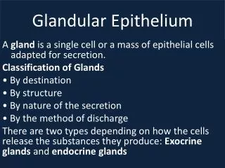
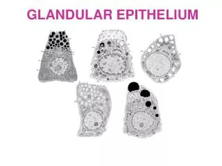
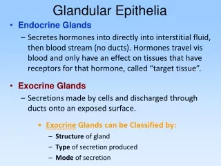
Glandular Epithelia • Endocrine Glands • Secretes hormones into directly into interstitial fluid, then blood stream (no ducts). Hormones travel vis blood and only have an effect on tissues that have receptors for that hormone, called “target tissue”. • Exocrine Glands • Secretions made by cells and discharged through ducts onto an exposed surface. • Exocrine Glands can be Classified by: • Structure of gland • Type of secretion produced • Mode of secretion
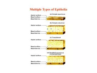
Structural Classification of Simple and Compound Exocrine Glands
Type of Secretion released from an Exocrine Gland Mucous – thick, sticky, viscous solution. Serous – watery, slippery, thin solution. Mixed – contains elements of both mucous and serous solutions.
Example of Mucous Glands The interior of the stomach is lined by a secretory sheet whose secretions protect the walls from acids and enzymes.
Example of Serous & Mixed Glands The submandibular salivary gland is a mixed gland containing cells that produce both serous and mucous secretions
Membranes = Epithelial Tissue Connective Tissue Membranes function to cover and protect body. • 4 types of Membranes • Mucous • Serous • Cutaneous • Synovial : )
MucousMembranes • Line passageways open to exterior. • Moistened by mucous (mucin + H2O). • 3 Layers: • Epithelium (simple or stratified) • Areolar CT called ‘Lamina Propria’ • Smooth Muscle called ‘Muscularis Mucosae’ • Roles: • Protection • Absorption • Secretion
Serous Membranes • Lines internal body cavities. • Thin, watery, serous fluid (transduate). • 2 layers: • Simple Squamous Epithelium (Mesothelium). • Thin areolar C.T. (subserous fascia). • Main function is to reduce friction. 1) Pleura, 2) Pericardium and 3) Peritoneum
Serous Membranes Internal body cavities are lined with membranes that produce serous secretions. Various deeper tissue Areolar C.T. Simple Squamous Epithelium
Modes of Secretion fromExocrine Glands • Merocrine (eccrine) Secretion • Exocytosis (vesicles fuse with PM and release contents from cell. • Most common mode of secretion. • Apocrine Secretion • Exocytosis but lipid rich content and can function as pheromones (‘scent glands’). • Holocrine Secretion • Involves entire cell, fills with vesicles and bursts, destroys the cell. Cell debris part of product.
Modes (or Mechanisms) of Secretion exposed surface
Mucous Serous Cutaneous Synovial
Fasciae Composed of Layers of fibrous C.T. in between various tissues. Roles: 1) Provides stability, maintains relative positions of organs. 2) Allows for distribution of blood, lymph vessels and nerves.
All organ systems are interconnected There are 3 types of Fasciae 1. Superficial Fascia 2. Deep Fascia 3. Subserous Fascia
Superficial Fascia(hypodermis) • Subcutaneous layer (adipose and dense irreg). • Insulation, padding, allows movement. • Deep Fascia • Dense connective tissue, lots of collagen. • Many layers with varying orientations. • Subserous Fascia • Areolar connective tissue. • In between deep fascia and serous membrane.