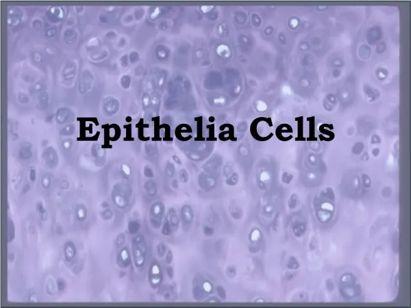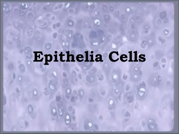
HISTOLOGY REVIEW Epithelia
350 likes | 1.37k Vues
HISTOLOGY REVIEW Epithelia. Dr. Tim Ballard Department of Biology and Marine Biology. Simple squamous epithelium. Kidney – median sagittal section – H&E – 10x objective.

HISTOLOGY REVIEW Epithelia
E N D
Presentation Transcript
HISTOLOGY REVIEWEpithelia Dr. Tim Ballard Department of Biology and Marine Biology
Simple squamous epithelium Kidney – median sagittal section – H&E – 10x objective Arrowheads indicate the locations of Bowman’s capsules within the cortex of the kidney. This is one place to locate simple squamous epithelium.
Simple squamous epithelium Kidney – median sagittal section – H&E – 40x objective lumen Bowman’s capsule Single layer of flattened cells with prominent flattened nucleus, very thin, lining the capsular space of Bowman’s capsule
Simple squamous epithelium Artery – cross section – H&E – 40xobjective lumen Flattened cells, one cell layer thick, lining the lumen of an artery
Simple cuboidal epithelium Kidney – median sagittal section – H&E – 10xobjective Arrowheads indicate renal tubules in the kidney cortex. “Tubule” is a key word, telling you this is one place to locate simple cuboidal epithelium.
Simple cuboidal epithelium Kidney – median sagittal section – H&E – 40xobjective lumen Approximate location of basement membrane lumen Approximate size of one cell Renal tubules (arrowheads) – note that the cells appear about as tall as they are wide and that there is a single layer of cells.
Simple cuboidal epithelium Kidney – median sagittal section – H&E – 40xobjective lumen In the renal medulla you find collecting ducts, lined with a single layer of simple cuboidal cells.
Simple cuboidal epithelium Thyroid gland – cross section – H&E – 40xobjective lumen lumen Simple cuboidal epithelium (arrowheads) forms follicles in the thyroid gland.
Simple cuboidal epithelium Pancreas – section – H&E – 40xobjective lumen Duct (arrowhead indicates location of the basement membrane) within the pancreas. Note the single layer of simple cuboidal cells.
Simple columnar epithelium Gallbladder – section – H&E – 10xobjective lumen Villi (arrowheads) – finger-like processes inside the gallbladder. This organ of the digestive system is line with simple columnar epithelium.
Simple columnar epithelium Gallbladder – section – H&E – 40xobjective Basement membrane would be located along this line. Approximate size of one cell lumen Note that the simple cuboidal cells are taller than they are wide.
Simple columnar epithelium Duodenum – section – H&E – 40xobjective Approximate size of one cell Basement membrane would be located along this line. lumen Although this looks different from the gallbladder, this is still simple columnar epithelium.
Ciliated pseudostratified columnar epithelium Trachea – cross section – H&E – 10xobjective lumen epithelium Basement membrane would be located along this line. Underlying connective tissue This is the signature epithelium of the respiratory system. Note the difference between epithelium above and connective tissue below the line.
Ciliated pseudostratified columnar epithelium Trachea – section – H&E – 40xobjective lumen cilia Basement membrane All cells contact the basement membrane, but not all cells reach the surface. It only appears stratified, hence the name.
Ciliated pseudostratified columnar epithelium Intrapulmonary bronchus – section – H&E – 40xobjective lumen cilia Basement membrane You can tell this isn’t stratified columnar epithelium because no can’t discern individual layers. Stratified columnar would have clear rows of nuclei.
Stratified squamous epithelium Esophagus – section – H&E – 10xobjective lumen How many layers of cells do you see here? Basement membrane Underlying connective tissue It is easy to see why this is called a stratified epithelium. I counted about 35 layers of cells stretching from the basement membrane to the apical surface.
(nonkeratinized) Stratified squamous epithelium Esophagus – section – H&E – 20xobjective lumen Basement membrane Cells at the basal surface are cuboidal and mitotic. As new cells are pushed to the top, they become increasingly squamous in shape.
Keratinized stratified squamous epithelium Human skin – section – H&E – 10xobjective Keratinized layers of cells free surface Basement membrane Again, look at the layers of cells between the basement membrane and the apical surface.
Keratinized stratified squamous epithelium Human skin – section – H&E – 40xobjective Keratinized layers of cells – very long, thin and flattened (scale-like) Free surface Epithelium Basal surface Cuboidal cells at the basal surface give way to increasingly flattened cells (squamous) in the upper layers. The cells are filled with water-proof keratin.
Stratified cuboidal epithelium Submandibular (salivary) gland – section – H&E – 40x objective Basal layer of cells Approximate size of one apical cell lumen Apical layer of cells Larger ducts of the body may have a stratified cuboidal epithelium, where the top layer of cells is cuboidal. There are usually only two layers of cells.
Stratified columnar epithelium Submandibular (salivary) gland – section – H&E – 40x objective lumen Apical layer of cells Approximate size of one apical cell What is the epithelial type? Basal layer of cells Very large ducts of the body may have a stratified columnar epithelium, where the top layer of cells is columnar. There are usually only two layers of cells.
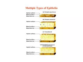
Transitional epithelium Ureter – cross section – H&E – 10x objective lumen Epithelial layer Underlying connective tissue layer Basal layer of cells Transitional epithelium is the signature epithelium of the urinary tract. It is found in the ureters and urinary bladder.
Transitional epithelium Ureter – cross section – H&E – 40x objective lumen Apical layer of cells Basal layer of cells When the organ walls are under low tension, the epithelium has 6 – 8 layers of cells. This is called transitional epithelium because the number of cell layers changes as the organ walls increase and decrease in tension.
Transitional epithelium Urinary bladder – section – H&E – 10x objective lumen Basement membrane The urinary bladder looks different from the ureter because it is a different organ with different function, but the epithelium is still transitional.
Transitional epithelium Urinary bladder – section – H&E – 40x objective Apical layer of cells lumen Basal layer of cells Basement membrane The distinguishing features of this epithelium are multiple layers and the very large “sofa pillow-like” cells at the apical surface. end