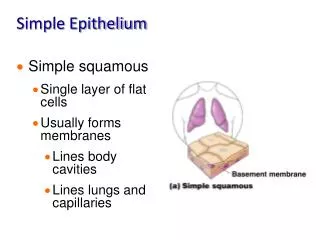
Glandular Epithelium
Glandular Epithelium. A gland is a single cell or a mass of epithelial cells adapted for secretion. Classification of Glands • By destination • By structure • By nature of the secretion • By the method of discharge

Glandular Epithelium
E N D
Presentation Transcript
Glandular Epithelium A gland is a single cell or a mass of epithelial cells adapted for secretion. Classification of Glands • By destination • By structure • By nature of the secretion • By the method of discharge There are two types depending on how the cells release the substances they produce: Exocrine glands and endocrine glands
Classification by Structure Secretorypart: unicellular / multicellular acinar (alveolar) / tubular coiled / branched Duct system: Simple gland (single duct) Complex gland (branched ducts) Branching ducts: Main > interlobular > Intralobular > Intercalary (ducts define structure of complex glands)
Exocrine glands secrete into ducts or directly onto a free surface. Their secretions include mucus, sweat, oil, ear wax and digestive enzymes. EXO = out side and crine = secret Structural classification of exocrine glands multicellular glands - most glands, have a distinctive appearance. Communicates with the surface
Examples include :- pancreas , stomach , sweat glands , salivary glands , mammary glands , sebaceous glands , etc . unicellular glands - single cell. goblet cells. No ducts .
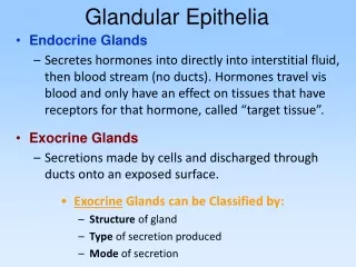
Endocrine glands - called “ductless glands” discharge their secretions into the intracellular fluid, where it diffuses into the blood stream. These secretions are hormones, or chemical messengers, which regulate many body functions.
Formation of glands from covering epithelia. During fetal development epithelial cells proliferate and penetrate the underlying connective tissue. They may-or may not-maintain a connection with the surface epithelium. When the connection is maintained, exocrine glands are formed; with the connection lost, endocrine glands are formed. Exocrine glands secrete to the body surface or gut via duct systems formed from the epithelial connection.
The cells of endocrine glands, which secrete hormone) can be arranged in cords or in follicles with lumens for storing the secretory product. From either the cords or follicles of endocrine cells, the secretory product is released outside the cells and picked up by the blood vessels for distribution throughout the body.
General structure of exocrine glands. Exocrine glands by definition have ducts that lead to an organ or body surface. Inside the gland the duct runs through connecting septa and branches repeatedly, until its smallest branches end in the secretory portions of the gland.
exocrine glands have a secretory portion , which contains the cells specialized for secretion , and ducts , which transport the secretion out of the gland .the morphology of these components allows the glands to be classified as follows :-
1-Ducts can be simple ( un branched ) or compound ( with two or more branches) 2-Secretory portions can be tubular ( either short or long and coiled ) or acinar ( round or globular) 3-Either type of secretory portion may be branched . 4-Compound glands can have tubular , acinar , or tubuloacinarsecretory portions
Classification by nature of secretionThe three major salivary glands: parotid, sub mandibular and sublingual.
Glands containing mucous acini (e.g. the sublingual glands) are called mucous glands. Glands containing serous acini (e.g. the parotid glands) are called serous glands. If both types of acini are present the gland is muco-serous or mixed gland .
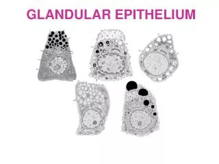
1- Mucous glands ( glands containing cells produce a viscous secretion that lubricates and or protects the inner lining of organs ) .Morphology : the cell is pyramidal in shape , flatten nuclei are displaced basally , their cytoplasm is completely filled with a light –staining , secretory product called mucus .
Serous glands ( glands with cell that produce watery secretions , which are often rich in enzymes ).Morphology : are similar in shape to mucous cells also pyramidal in shape , spherical or round nuclei are displaced basally by secretory granules that accumulate in the apical regions .
Mixed glands ( glands that contain a mixture of both mucous and serous secretory cells). Morphology : mucous cells predominant , serous cells form a crescent or moon-shaped cap over the mucous cells , called serous demilune
Classification by method of secretion• Merocrine• Apocrine• Holocrine
Merocrine secretion is exocytosis• Membrane bounded component approaches cell surface• It fuses with plasma membrane• Its contents are in continuity with the extracellular space• Plasma membrane transiently larger• Membrane retrieved, stabilizing cell surface area
Apocrine secretion• Non-membrane bounded structure (e.g. lipid) approachescell surface• Makes contact and pushes up apical membrane• Thin layer of apical cytoplasm drapes around droplet• Membrane surrounding drop[let pinches off from cell• Plasma membrane transiently smaller• Membrane added to regain original area
Holocrine secretion• Disintegration of the cell• Release of contents• Discharge of whole cell
Endocytosis• Engulfing material initially outside the cell• Opposite of exocytosis (merocrine secretion)• Endo- & Exo-cytosis are coupled in transepithelial transportTransepithelial Transport• material endocytosed at one surface• transport vesicle shuttles across cytoplasm• material exocytosed at opposite surface
Golgi Apparatus:Structure• Stack of disc-shaped cisternae• One side of discs are flattened; other concave• Discs have swellings at their edges• Distal swellings pinch off as migratory Golgi Vacuoles
Golgi Apparatus:Function• Packaging through condensation of contents• Transport• Adding sugars to proteins and lipids (Glycosylation)Golgi Product Destinations• Majority extruded in secretory vesicles• Some retained for use in the cells (e.g. lysosomes)• Some enters the plasma membrane (Glycocalyx)
Glycosylation & Specificity• Branching sugars offer complex shapes for specificinteractions in the glycocalyx• Destruction of this layer by enzymes alters manyspecificity based properties of cells:- adhesion to substrates & neighboring cells- mobility of cells- communication with neighboring cells- contact inhibition of movement and division
Control of Secretion • Nervous • Endocrine control • Neuro-endocrine control • Negative feedback chemical mechanism
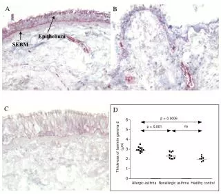
An islet of Langerhans surrounded by exocrine pancreatic acini (high power).
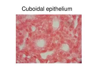
Simple cuboidal epithelium of the thyroid gland surrounding homogeneous colloid in each follicle.