EPITHELIUM
EPITHELIUM. What is meant by epithelium? Define epithelium Why epithelium is called epithelium Why epithelium is given this name. Following statements regarding epithelium are correct except


EPITHELIUM
E N D
Presentation Transcript
What is meant by epithelium? • Define epithelium • Why epithelium is called epithelium • Why epithelium is given this name
Following statements regarding epithelium are correct except Epithelium is layered collection of cells covering external and internal surfaces of the body, including the lining of vessels and other small and large cavities. The term, epithelium was used to refer to the cellular covering of the nipple. Epithelium is predominantly cellular tissue with numerous intercellular junctions Epithelium is a layered collection of cells with large amount of intercellular material. The main function of the epithelial lining of gastro-intestinal tract is absorption.
Epithelium, pl. epithelia. [Gr. epi- on + Gr. thēlē nipple] • Epithelium is layered collection of cells covering external and internal surfaces of the body, including the lining of vessels and other small and large cavities.
This term (epithelium) was used to refer to the cellular covering of the nipple. • Nipple is also called thelium. • Thelium is a papilla or a nipple. • Later on the term epithelium has been extended to include the cellular coverings of the external and internal surfaces of the body.
The term epithelium derives from the fact that the interface between epithelium and the underlying connective tissue frequently shows projections and depressions (epidermal and dermal papillae) which appear as small nipples.
Epithelium is predominantly cellular tissue with numerous intercellular junctions and very little intercellular substance. • Epithelium is a layered collection of cells, held together in close contact by intercellular junctions, with a small amount of intercellular material.
Epithelium is the covering of internal and external surfaces of the body, including the lining of vessels and other small and large cavities. • Simple Squamous Epithelium lining a Capillary wall. • H&E stain. Magnification 400x.
Functions of epithelial tissue are protection, secretion, absorption and sensation.
How the epithelium is separated from the underlying connective tissue
ALLAH THE KHALLAQ-UL-ALLEEM خلاق العليم has placed a membrane between the epithelium and connective tissue called basement membrane. • Name the components of basement membrane
This membrane consists of two components, basal lamina derived from epithelium and reticular lamina derived from connective tissue. • Describe the blood supply of epithelium • How the epithelium is gets its nutrition
Epithelium is avascular. • Nutritive materials and oxygen enter it by diffusion through the cells and across basal lamina.
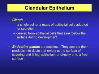
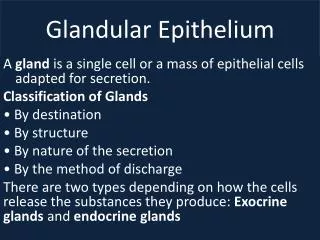
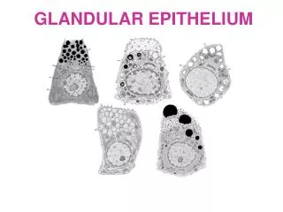
Classification of Epithelium • There are two types of epithelial tissue: • Surface Epithelium • Glandular Epithelium
SURFACE EPITHELIUM • The epithelium covering the body or lining the surfaces of body cavities, viscera and tubes or hollow channels in the body is called surface epithelium. • It is divided into two main varieties.
Classical: Here the cells forming the epithelium are arranged in regular layers. It may be single layered or many layered. • Non-Classical: This type of epithelium contains single layered as well as many layered cells. They are not regular in arrangement.
Classical Epithelium • Unilayered epithelium • Simple squamous • Simple cuboidal • Simple columnar
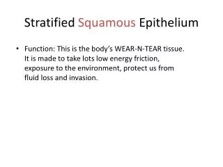
Classical Epithelium • Multilayered epithelium • Stratified squamous • Stratified cuboidal • Stratified columnar
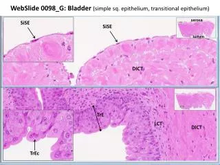
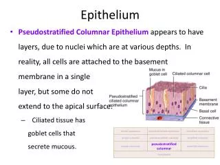
Non-Classical epithelium • Complex Epithelium • Pseudostratified epithelium • Transitional epithelium • Seminiferous epithelium
Non-Classical epithelium • Specialized Sensory Epithelium • Olfactory epithelium • Gustatory epithelium • Auditory epithelium • Retinal epithelium
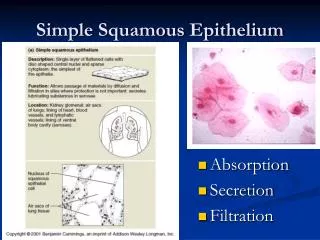
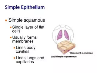
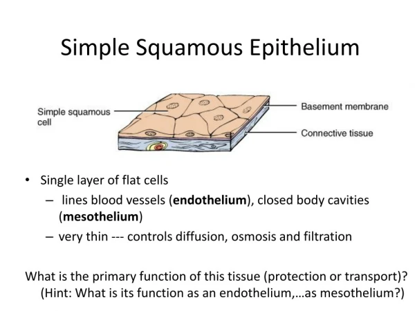
Simple Squamous Epithelium • Simple squamous epithelium consists of a single layer of flat cells that resemble the tiles on a floor. • The nucleus of each cell is centrally placed and is rounded or oval.
Simple Squamous Epithelium lining a Capillary wall. • H&E stain. Magnification 400x. • The lining of all blood vessels is simple squamous epithelium, and usually is referred to as the endothelium.
Simple Squamous Epithelium lining the blood vessels called Vascular endothelium. • Mallory's stain. Magnification 400x. • Note the pointer indicating the simple squamous cells of vascular endothelium.
Simple Squamous Epithelium. Bowman's capsule, kidney glomerulus. • Stain hematoxylin and eosin (H&E). Magnification 400x. • The pointer indicates the thin simple squamous epithelium, composed of a single layer of flattened cells often showing conspicuous bulging nuclei.
Simple Squamous Epithelium of Bowman's capsule. • H&E stain. Magnification 600x. • At increased magnification note the delicate thin cytoplasm of the simple squamous epithelial cells and the presence of prominent ovoid nuclei.
Simple Squamous Epithelium of Bowman's capsule. • Mallory's stain. Magnification 400x. • Here again you see the thin flattened simple squamous epithelial membrane with its characteristic structure.
SITES: Endodermal: Alveoli of lungs;
Mesodermal: Renal Corpuscles and thin segments of Loope of HenIe of kidneys; Lining of the Cardiovascular system (Heart, Blood vessels and Lymphatics); Lining of Body Cavities (Pleural, Pericardial and Peritoneal). Subarachnoid and Subdural cavities Chambers of the Eye Perilymphatic spaces of the Ear Lining of the synovial membranes of Joint cavity and Bursae
The surface of the ovary is covered, in the young female, with a layer of cuboidal cells which become flattened later in life. • The flat, thin cells are ideally suited to perform the functions of diffusion (alveoli of lungs) and filtration (glomeruli of kidneys).
Simple squamous epithelium lining the cardiovascular system is given the special name of endothelium. • This is a wrong name given to this epithelium because it does not develop from endoderm rather it develops from mesoderm.
The epithelial lining of body cavities is called mesothelium, • It does develop from mesoderm but many other epithelia develop from mesoderm yet they are not given the name mesothelium,
The epithelium lining the synovial membranes of joint cavities and bursae, chambers of the eye, perilymphatic spaces of the ear, subarachnoid and subdural cavities is sometimes called mesenchymal epithelium which is a formality and creates inconvenience.
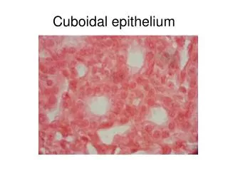
Simple Cuboidal Epithelium • Simple cuboidal epithelium consists of a single layer of cells shaped like cubes. • The nucleus of each cell is rounded and centrally placed.
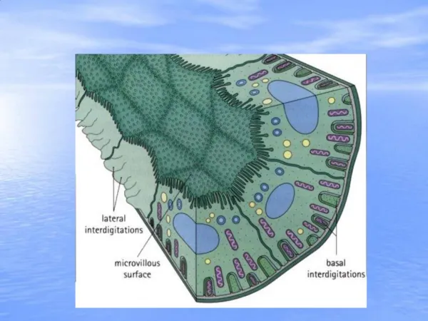
Simple Cuboidal Epithelium in Renal (kidney) tubules. H&E stain. Magnification 400x. Simple cuboidal epithelium consists of a single layer of cells that in sections appear to be as wide as they are tall. In this frame, the upper pointer locates the supporting basal membrane for the simple cuboidal epithelium, while the lower pointer indicates a structure called the brush border. TEM shows that the brush border is composed of structures called microvilli.
TEM [Tomographic Electron Micrograph] • Tomo-graphic [Gr. tomē a cutting + graphein to write] • An x-ray apparatus which makes a roentgenogram of a layer of tissue at any depth. • Roentgenogram is a film produced by roentgenography.
Roentgenography is the photography by means of roentgen rays. Special techniques for roentgenography of different areas of the body have been given specific names, such as angiography, angiocardiography, encephalography, portography, pyelography, etc.
Roentgen was a German physicist. His name was Wilhelm Conrad Röntgen. He was born in 1845 and died in 1923. He discovered roentgen rays in 1895. He was the winner of the Nobel prize in physics for 1901.