
Uploaded by
orpah
15 SLIDES
359 VUES
150LIKES
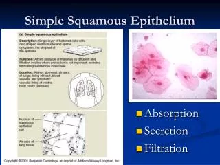
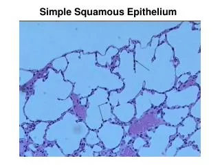
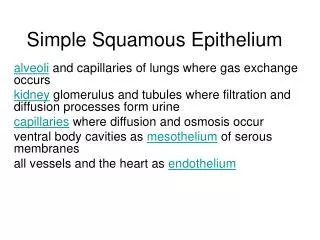
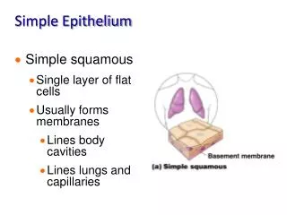
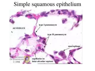
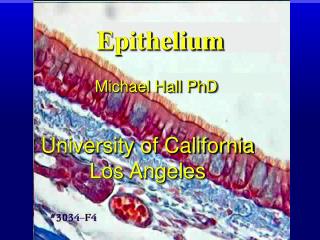
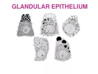
Squamous Epithelium
DESCRIPTION
Tissue Slides PowerPoint BIO 138 Mr. Hauser Use these images as a resource to compare what you are observing with the microscopes. Squamous Epithelium. Simple Cuboidal Epithelium. Simple Columnar Epithelium. Stratified Squamous Epithelium. Stratified Cuboidal Epithelium.
Download
1 / 15
Download Presentation

Télécharger la présentation
Squamous Epithelium
An Image/Link below is provided (as is) to download presentation
Download Policy: Content on the Website is provided to you AS IS for your information and personal use and may not be sold / licensed / shared on other websites without getting consent from its author.
Content is provided to you AS IS for your information and personal use only.
Download presentation by click this link.
While downloading, if for some reason you are not able to download a presentation, the publisher may have deleted the file from their server.
During download, if you can't get a presentation, the file might be deleted by the publisher.
E N D
Presentation Transcript
Tissue Slides PowerPointBIO 138Mr. HauserUse these images as a resource to compare what you are observing with the microscopes.
Cartilage (Elastic) Cartilage (Hyaline)
Skeletal Muscle Tissue (striated)
Nervous Tissue What is the dark dot in the cell body of the neuron? What are the ‘branches’ coming out of the cell body of the neuron called?


More Related
Audio
Live Player