The cardiovascular system
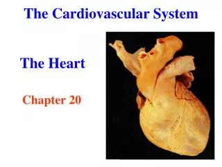
The cardiovascular system. Mary Chiang B8. The parts of the cardiovascular system. Heart Veins Arteries Capillaries. The outside of the heart. Aorta Pulmonary artery Pulmonary vein Coronary arteries Vena cava (superior) Vena cava (inferior). 1. 5. 2. 3. 4. 4. 4. 4. 6.

The cardiovascular system
E N D
Presentation Transcript
The cardiovascular system Mary Chiang B8
The parts of the cardiovascular system • Heart • Veins • Arteries • Capillaries
The outside of the heart Aorta Pulmonary artery Pulmonary vein Coronary arteries Vena cava(superior) Vena cava (inferior) 1 5 2 3 4 4 4 4 6
What do they do? • Aorta • the large artery in charge of allocating blood rich in oxygen to the body • Pulmonary artery • The artery that sends oxygen-less blood from the right ventricle to the lungs • Pulmonary vein • From the lung, oxygen-rich blood travels in this vein to the left atrium • Coronary arteries • Give the heart oxygen-rich blood so it stays healthy and in working condition; Two branches of these coronary arteries send the oxygen-rich blood to where it needs to be in the heart • Vena cava (superior) • Oxygen-less blood from the upper parts of the body (head, neck, and arms) travels in this vein to the right atrium • Vena cava (inferior) • Oxygen-less blood from the lower parts of the body (legs, etc) is pumped through this vein to the right atrium
The inside of the heart Left atrium Right atrium Left ventricle Right ventricle Valves Septum 1 5 5 5 2 3 5 4 6
What do they do? • Left atrium • Pulmonary veins bring oxygen-rich blood to this chamber; then the blood goes on to the left ventricle • Right atrium • Vena cava sends oxygen-less blood to this chamber; then the blood goes to the right ventricle • Left ventricle • Left atrium, oxygen-rich blood comes here and then to the aorta • Right ventricle • Oxygen-less blood from the right atrium comes to this chamber and then to the pulmonary artery • Valves • Ensuring the correct flow of blood throughout the heart and the body, these “biological gateways” are very important to the heart and the body • Septum • Large mass of muscle that separates the left and right side of the heart
Purpose of the circulatory system • Without hearts, humans would die. Drop dead. That’s the sound of them falling dead to the floor without their hearts. • Without the heart and the veins and arteries, detectives and other crime-solving bosses could not dramatically say, “He’s dead” because there would not a pulse for them to feel for. • The pumping of the heart ensures blood flow which distributes oxygen received from respiration and other much-needed nutrients received from digestion to the rest of the body. • Blood also carries the immune system cells to watch out for infections, and it brings waste to the kidneys and liver to be filtered and excreted out of the body. PLOP.
Journey to the center of the heart (and lungs) PARTS OF THE BODY O O O O O O OTHER PARTS OF THE BODY
FEEL YOUR HEARTBEAT • *Cue the The Fray song “Heartbeat”* • Anyway, your heartbeat is caused when the upper chambers contract to send blood to the corresponding lower chambers • To make the heart , an “electrical impulse” is generated by nodes, special cells on the heart • Once the heart contracts, the blood from the upper chambers (WHICH ARE CALLED?) flows into the lower chambers (WHICH ARE CALLED?) through the valves (Everyone remember what those are????) • To send the blood out of the heart and into the body, the nodes work their stuff again, sending out those handy-dandy “electrical impulses” that make the ventricles contract, squeezing the blood out so that it can make its way to the lungs and the body contract
LUB-DUB • What makes that famous LUB sound in the “lub-dub” heart beat? After the blood is squeezed through them, they snap closed so the blood does not flow back in the heart • And what about DUB, you say? Well, those valves between the ventricles and the rest of the body snap shut. More blood enters the atria and EVERYTHING BEGINS ANON. VALVES!!!!!!
What’s blood? • Blood is a tissue of the body in liquid form • The watery portion of blood is plasma which makes up 55% of the blood volume. The 45% of the blood is made up of the blood cells: • Red blood cells • White blood cells • Platelets • carries oxygen to the heart • takes away carbon dioxide from body tissue to the lungs • essential to humans, cleaning and nourishing the physical bodies to keep us alive
Arteries • carry oxygen-rich blood away from the heart • rich red color • thick walled, excluding the cranium and vertebral canals which are thin-walled • The size and type of an artery depend on their function • large, elastic arteries, like the aorta • small, muscular arteries, like the temporal or radial arteries • Inner coat • Tunica intima • Middle coat • Tunica media • Outer coat (strongest coat) • Tunica adventitia
veins • Vessels that carry blood away from peripheral tissues and toward the heart(non-oxygenated blood) • Blue in color • Thin walled • Valves control the “unidirectional” flow of the blood (sometimes against gravity) • Same 3 layers as arteries but not as well defined with poor muscle and tissue content
Capillaries • Capillaries and tissues exchange nutrients and metabolites • Networks of microscopic endothelial tubes b/w the metarterioles and venules • There are two types of capillaries: • Fenestrated: LARGE molecules may pass over the walls • Examples: • endocrine glands • pancreas • Continuous: small molecules pass over walls • Examples: • Skin • Connective tissue • Muscles • Roughly around 6-8 micron, barely wide enough for cells to file through in single file; however, their size does differ • Small in brain and intestines • Large in skin and bone marrow • The interstitial fluid and the blood constantly exchange gases, nutrients, metabolites, and water at the enormous capillary bed
Homeostasis • The cardiovascular system plays such an important role in homeostasis that the cardiovascular system develops in an organism during the fourth week after fertilization before any major organ systems develop • To maintain a favorable cellular environment, the body depends on the constant circulation of blood throughout the body • The blood must reach the thousands and thousands of capillaries in every single part of the body, whether it be a single cell or a whole organ • The capillary blood carries essential materials and passes the important nutrients and materials into the cellular fluid and removes waste products
Evolutionary History of the Cardiovascular system • Invertebrate hearts • Simple • Open circulatory system w/ no actual blood vessels or very few blood vessels • Blood pumped through tissues then back to pumping mechanism • No actual heart • Fish hearts (2 chambers: atrium and ventricle) • Closed circulatory system • One large vessel pumps blood to give fish gills oxygen and to the rest of the body • Frog hearts (3 chambered heart: Two atria, one ventricle) • Link between land animals and water animals • Oxygen-rich and oxygen-less blood separate in the heart • Turtle hearts (3 ½ chambered heart) • Partly-formed Septum in heart • Blood mixes, but timing of ventricle pumping minimizes chance that the oxygen-rich and oxygen-less blood will mix together • Human hearts (4 chambered: Two atria, two ventricles) • Fully formed septum
Works Cited The Franklin Institute (2013). Blood: White Blood Cells - The Human Heart: An Online Exploration from The Franklin Institute, made possible by Unisys. Retrieved March 31, 2013, from http://www.fi.edu/learn/heart/blood/white.html National Cancer Institute (2012, November 27). SEER Training: Introduction to the Cardiovascular System. Retrieved March 31, 2013, from http://training.seer.cancer.gov/anatomy/cardiovascular/ National Geographic (n.d.). Heart, Heart Information, Cardiovascular Facts, News, Photos -- National Geographic. Retrieved March 31, 2013, from http://science.nationalgeographic.com/science/health-and-human-body/human-body/heart-article.html OCAL. (2009, May 11). Lungs clip art. Retrieved April 2, 2013, from http://www.clker.com/clipart-lungs-1.html Scouville, H. (n.d.). Evolution of the Human Heart. Retrieved March 31, 2013, from http://evolution.about.com/od/humans/a/Evolution-Of-The-Human-Heart.htm Iqbal, A. (2010). Arteries. Retrieved March 31, 2013, from http://www.mananatomy.com/basic-anatomy/arteries Iqbal, A. (2010). Capillaries. Retrieved March 31, 2013, from http://www.mananatomy.com/basic-anatomy/capillaries Iqbal, A. (2011). Components of Cardiovascular System (CVS). Retrieved March 31, 2013, from http://www.mananatomy.com/basic-anatomy/components-cardiovascular-system-cvs Iqbal, A. (2010). Veins. Retrieved March 31, 2013, from http://www.mananatomy.com/basic-anatomy/veins