Brain – Short Version
12. P A R T A. Brain – Short Version. Central Nervous System (CNS). CNS – composed of the brain and spinal cord Cephalization Elaboration of the anterior portion of the CNS Increase in number of neurons in the head Highest level is reached in the human brain. The Brain.

Brain – Short Version
E N D
Presentation Transcript
12 P A R T A Brain – Short Version
Central Nervous System (CNS) • CNS – composed of the brain and spinal cord • Cephalization • Elaboration of the anterior portion of the CNS • Increase in number of neurons in the head • Highest level is reached in the human brain
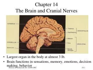
The Brain • Composed of an outer layer of wrinkled, pinkish gray tissue (gray matter) – that is composed of non-myelinated neurons and an inner core of whitish tissue (white matter) composed of myelinated neurons. • Surface anatomy includes cerebral hemispheres, cerebellum, and brain stem
Embryonic Development • During the first 26 days of development: • Ectoderm thickens forming the neural plate • The neural plate invaginates, forming the neural groove • The neural groove fuses dorsally and forms the neural tube
Embryonic Development Figure 12.1
Primary Brain Vesicles • The anterior end of the neural tube expands and constricts to form the three primary brain vesicles • Prosencephalon – the forebrain • Mesencephalon – the midbrain • Rhombencephalon – hindbrain
Neural Tube and Primary Brain Vesicles Figure 12.2a, b
Secondary Brain Vesicles Figure 12.2c
Secondary Brain Vesicles • In week 5 of embryonic development, secondary brain vesicles form • Telencephalon and diencephalon arise from the forebrain • Mesencephalon remains undivided • Metencephalon and myelencephalon arise from the hindbrain
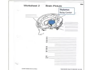
Adult Brain Structures • Fates of the secondary brain vesicles: • Telencephalon – cerebrum: cortex, white matter, and basal nuclei • Diencephalon – thalamus, hypothalamus, and epithalamus • Mesencephalon – brain stem: midbrain • Metencephalon – brain stem: pons • Myelencephalon – brain stem: medulla oblongata
Adult Neural Canal Regions Figure 12.2c, d
Adult Neural Canal Regions • Adult structures derived from the neural canal • Telencephalon – lateral ventricles • Diencephalon – third ventricle • Mesencephalon – cerebral aqueduct • Metencephalon and myelencephalon – fourth ventricle
Adult Neural Canal Regions Figure 12.2c, e
Space Restriction and Brain Development Figure 12.3
Basic Pattern of the Central Nervous System • Spinal Cord (two layers – outer white matter and inner gray) • Central cavity surrounded by a gray matter core • External to which is white matter composed of myelinated fiber tracts • Brain (three layers outer gray – mid white then an inner area of gray matter) • Similar to spinal cord but with additional areas of gray matter • Cerebellum has gray matter in nuclei • Cerebrum has nuclei and additional gray matter in the cortex
Basic Pattern of the Central Nervous System Figure 12.4
Meninges Figure 12.24a
Dura Mater • Leathery, strong meninx composed of two fibrous connective tissue layers • The two layers separate in certain areas and form dural sinuses
Dura Mater • Three dural septa extend inward and limit excessive movement of the brain • Falx cerebri – fold that dips into the longitudinal fissure • Falx cerebelli – runs along the vermis of the cerebellum • Tentorium cerebelli – horizontal dural fold extends into the transverse fissure
Dura Mater Figure 12.25
Arachnoid Mater • The middle meninx, which forms a loose brain covering • It is separated from the dura mater by the subdural space • Beneath the arachnoid is a wide subarachnoid space filled with CSF and large blood vessels • Arachnoid villi protrude superiorly and permit CSF to be absorbed into venous blood
Arachnoid Mater Figure 12.24a
Pia Mater • Deep meninx composed of delicate connective tissue that clings tightly to the brain
Ventricles of the Brain • Arise from expansion of the lumen of the neural tube • The ventricles are: • The paired C-shaped lateral ventricles • The third ventricle found in the diencephalon • The fourth ventricle found in the hindbrain dorsal to the pons
Ventricles of the Brain Figure 12.5
Cerebrospinal Fluid (CSF) • Watery solution similar in composition to blood plasma • Contains less protein and different ion concentrations than plasma • Forms a liquid cushion that gives buoyancy to the CNS organs
Cerebrospinal Fluid (CSF) • Prevents the brain from crushing under its own weight • Protects the CNS from blows and other trauma • Nourishes the brain and carries chemical signals throughout it
Circulation of CSF Figure 12.26b
Choroid Plexuses • Clusters of capillaries that form tissue fluid filters, which hang from the roof of each ventricle • Have ion pumps that allow them to alter ion concentrations of the CSF • Help cleanse CSF by removing wastes
Choroid Plexuses Figure 12.26a
Blood-Brain Barrier • Protective mechanism that helps maintain a stable environment for the brain • Bloodborne substances are separated from neurons by: • Continuous endothelium of capillary walls • Relatively thick basal lamina • Bulbous feet of astrocytes
Blood-Brain Barrier: Functions • Selective barrier that allows nutrients to pass freely • Is ineffective against substances that can diffuse through plasma membranes • Absent in some areas (vomiting center and the hypothalamus), allowing these areas to monitor the chemical composition of the blood • Stress increases the ability of chemicals to pass through the blood-brain barrier
Cerebrum composed of the Cerebral Hemispheres • Form the superior part of the brain and make up 83% of its mass • Contain ridges (gyri) and shallow grooves (sulci) • Contain deep grooves called fissures • Are separated by the longitudinal fissure • Have three basic regions: cortex, white matter, and basal nuclei
Major Lobes, Gyri, and Sulci of the Cerebral Hemisphere • Deep sulci divide the hemispheres into five lobes: • Frontal, parietal, temporal, occipital, and insula • Central sulcus – separates the frontal and parietal lobes
Brain Lobes Figure 12.6a–b
Major Lobes, Gyri, and Sulci of the Cerebral Hemisphere • Parieto-occipital sulcus – separates the parietal and occipital lobes • Lateral sulcus – separates the parietal and temporal lobes • The precentral and postcentral gyri border the central sulcus
Cerebral Cortex • The cortex – superficial gray matter; accounts for 40% of the mass of the brain • It enables sensation, communication, memory, understanding, and voluntary movements • Each hemisphere acts contralaterally (controls the opposite side of the body) • Hemispheres are not equal in function (right more artistic – left more technical) • No functional area acts alone; conscious behavior involves the entire cortex
Functional Areas of the Cerebral Cortex • The three types of functional areas are: • Motor areas – control voluntary movement • Sensory areas – conscious awareness of sensation • Association areas – integrate diverse information • Several anatomists attempted to find physiologic (functional) areas of the – K. Broadman in 1906 was the best. He found 52 functional areas – thus numbered. However, now we no that one area of the brain functionally does it all for a function. It is a combination of several areas confluently working together.
Functional Areas of the Cerebral Cortex Figure 12.8a
Functional Areas of the Cerebral Cortex Figure 12.8b
Cerebral Cortex: Motor Areas • Primary (somatic) motor cortex (Broadman 4) • Premotor cortex (Broadman 6) • Broca’s area (Speech area) (Broadman 45 and 44) • Frontal eye field (Broadman 8)
Primary Motor Cortex • Located in the precentral gyrus • Pyramidal cells whose axons make up the corticospinal tracts • Allows conscious control of precise, skilled, voluntary movements
Primary Motor Cortex Homunculus Figure 12.9.1
Premotor Cortex • Located anterior to the precentral gyrus • Controls learned, repetitious, or patterned motor skills • Coordinates simultaneous or sequential actions • Involved in the planning of movements
Broca’s Area • Broca’s area • Located anterior to the inferior region of the premotor area • Present in one hemisphere (usually the left) • A motor speech area that directs muscles of the tongue • Is active as one prepares to speak
Frontal Eye Field • Frontal eye field • Located anterior to the premotor cortex and superior to Broca’s area • Controls voluntary eye movement • Note: vision itself is in the occipital lobe
Sensory Areas • Primary somatosensory cortex (1, 2, 3) • Somatosensory association cortex (5, 7) • Visual (19, 18, 17) and auditory areas ( 41, 42, 22) • Olfactory Cortex (34, 28) • Gustatory Cortex (Insula) • Visceral Cortex (Insula) • Vestibular Cortex (Insula)
Sensory Areas Figure 12.8a
Functional Areas of the Cerebral Cortex Figure 12.8b
PrImary Somatosensory Cortex • Located in the postcentral gyrus, this area: • Receives information from the skin and skeletal muscles • Exhibits spatial discrimination