Download
1 / 35
490 likes | 2.52k Vues
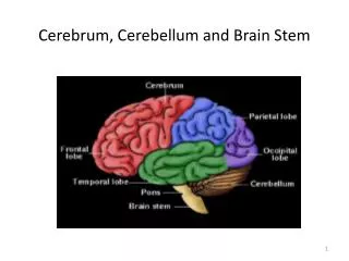
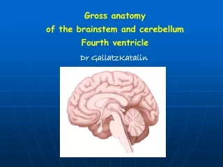
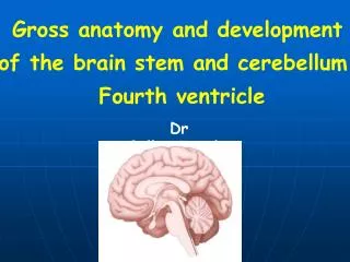
Gross anatomy and development of the brain stem and cerebellum. Fourth ventricle. Dr GallatzKatalin. The brain. Telencephalon Diencephalon Cerebellum Brain stem . Parts of the brain stem. Mesencephalon Pons Medulla. Ventral surface of the hypothalamus and brainstem.

E N D
Gross anatomy and development of the brain stem and cerebellum. Fourth ventricle Dr GallatzKatalin
The brain • Telencephalon • Diencephalon • Cerebellum • Brain stem
Parts of the brain stem • Mesencephalon • Pons • Medulla
Ventral surface of the hypothalamus and brainstem 1. cerebral peduncle 2. interpeduncular fossa 3. oculomotor nerve 4. optic tract 5. pons 4 5 5 3 1 2 4 5
Dorsal surface of the diencephalon and brainstem sc – superior colliculus, ic – inferior colliculus, scp – sup. cerebellar peduncle th sc ic scp pons medulla
Brain Stem Midbrain Cranial Nerve III., IV. • pupillary reflex • eye movements
The midbrain or mesencephalon (from the Greek mesos – middle, and enkephalos) - is associated with vision, hearing, motor control, pain control sleep/wake, and temperature regulation
Parts of the mesencephalon • - tectum • inferior colliculi • superior colliculi • - cerebral peduncle • tegmentum • crus cerebri
Midbrain Ventral surface • Crus cerebri (1) • Interpeduncular fossa (2) oculomotor nerves(3) • Posterior perforated substance 3 1 2
Midbrain MGB Dorsal surface • Superior colliculus(1) constitute centers for visual reflexes • Inferior colliculus(2) associated with auditory pathway • Brachium of superiorcolliculus LGB • Brachium of inferior colliculus MGB LGB 1 trochlear n. 2 scp
Brain Stem • Pons • Cranial Nerves V., VI., VII.
Parts of the pons • tegmentum – dorsal surface • base –venral surface
pontine tegmentum base
Pons Ventral surface • Basilar sulcus • Pontomedullary junction: from medial to lateral, abducent, facial and vestibulocochlear nerves Middle cerebellar peduncle • Trigeminal nerve • Pontocerebellar trigone: the junction of medulla, pons and cerebellum
Pons Dorsal surface • Medial eminence(1) • Facial colliculus(2) • Locus ceruleus(3) 1 3 2
Brain Stem • Medulla Cranial Nerves IX, X, XI, XII • Pharyngeal (Gag) Reflex • Tracheal (Cough) Reflex • Cardiovascular, respiratory centers
Parts of the medulla • opened part – rostral part • closed part – caudal part dorsal surface ventral surface
Medulla oblongata ventral surface • Pyramid: contain pyramidal tract(corticospinal tract) • Decussation of pyramid: formed by crossing fibers of corticospinal tract • Olive: inferior olivary nucleus • Medial parolivary sulcus hypoglossal nerve Lat. parolivary sulcus:glossopharyngeal, vagus accessory nerves
Medulla oblongata Dorsal surface • Caudal portion • Gracile tubercle: produced by underlying gracile nucleus • Cuneate tubercle: marks the site of cuneate nucleus • Inferior cerebellar peduncle • Obex • Rostral portion: • forms the caudal half of rhomboid fossa
Fourth ventricle Position • Situated ventral to the cerebellum, and dorsal to the pons and cranial half of medulla
Borders of the rhomboid fossa – floor of the fourth ventricle • Inferolateral: inferior cerebellar peduncle • Superolateral: superior cerebellar peduncle • Lateral recess
Rhomboid fossa – floor of the fourth ventricle • Facial colliculus, medial eminence • Hypoglossal trianglehypoglossal nucleus • Vagal triangle: dorsal nucleus of vagusnerve • Funiculus separans • Area postrema • Locus ceruleus
Roof of the fourth ventricle • Anterior part: formed by superior cerebellar peduncle and superior medullary velum • Posterior part: formed by inferior medullary velum,tela choroidea and choroid plexus of fourth ventricle • Three apertures Median aperture(Magendi) Two lateral apertures(Luschka)
Position of the cerebellum • It lies in the posterior cranial fossa • The tentorium cerebelli separates it from the occipital lobe of the cerebrum
Cerebellar peduncles - Inferior cerebellar peduncle(1)connects it with medulla - Middle cerebellar peduncle(3)connects it with pons, - Superior cerebellar peduncle(2)-connects it with midbrain, 1 2 3
External features Consists of twocerebellar hemispheres united in the midline by the vermis
Lobes and fissures of the cerebellum • Two deep fissures • Primary fissure • Posterolateral fissure • Horizontal fissure • Three lobes • Flocculonodular lobe flocculus and nodule • Anterior lobe • Posterior lobe
Lobs Anterior lobe Primary fissure Posterior lobe Flocculonodular lobe Posterolateral fissure
Internal structures Cerebellar nuclei Fastigial nucleus Globose nucleus Dentate nucleus Emboliform nucleus Cerebellar cortex
Functional divisions • Vestibulocerebellum • Archicerebellum • Flocculonodular lobe • Spinocerebellum • Paleocerebellum • Vermis and intermediate zone • Cerebrocerebellum • Neocerebellum • Lateral zone Intermediate zone Vermis Lateral zone Flocculonodular lobe