Download
1 / 39
390 likes | 505 Vues
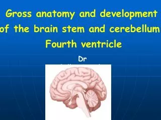
Gross anatomy of the brainstem and cerebellum Fourth ventricle. Dr GallatzKatalin. The brain. Telencephalon Diencephalon Cerebellum Brainstem. Parts of the brainstem. Mesencephalon Pons Medulla. Ventral surface of the hypothalamus and brainstem. 1. cerebral peduncle

E N D
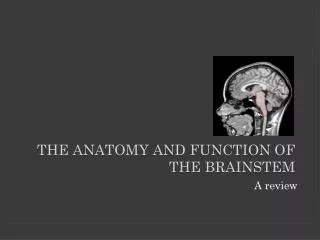
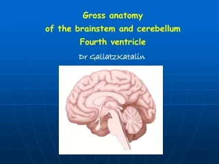
Gross anatomy of the brainstem and cerebellum Fourth ventricle Dr GallatzKatalin
The brain • Telencephalon • Diencephalon • Cerebellum • Brainstem
Parts of thebrainstem • Mesencephalon • Pons • Medulla
Ventral surface of the hypothalamus and brainstem 1. cerebral peduncle 2. interpeduncular fossa 3. optic tract 3 1 2 pons 5 medulla 5
Dorsal surface of the diencephalon and brainstem sc – superior colliculus, ic – inferior colliculus, scp – sup. cerebellar peduncle thalamus sc ic scp pons medulla
Brain Stem Midbrain Cranial Nerve III., IV. • pupillary reflex • eye movements
The midbrain or mesencephalon (from the Greek mesos – middle, and enkephalos) - is associated with vision, hearing, motor control, pain control, sleep/wake, and temperature regulation
Parts of themesencephalon TECTUM inferiorcolliculi superiorcolliculi CEREBRAL PEDUNCLE tegmentum cruscerebri TECTUM TEGMENTUM
Midbrain - Mesencephalon Ventral surface • Cerebralpeduncle(1) • Interpeduncular fossa (2) oculomotor nerves(3) • Posterior perforated substance 3 2 1
Midbrain - Mesencephalon trochlear n. Dorsal surface • Superior colliculus(1) centers for visual reflexes • Inferior colliculus(2) • associated with auditory pathway • Brachium of superiorcolliculus LGB • Brachium of inferior colliculus MGB scp
Brainstem PONS Cranial nerves V., VI., VII.
Parts of the pons tegmentum – dorsal surface base –venral surface
pontine tegmentum base
Pons Ventral surface • Basilar sulcus • Pontomedullaryjunction: from medial to lateral, VI, VII. VIII nerves Middle cerebellar peduncle • Trigeminal nerve • Pontocerebellar trigone: the junction of medulla, pons and cerebellum
Pons Dorsal surface – rostral part of therhomboid fossa part • Medialeminence(1) • Facialcolliculus(2) • Locusceruleus(3) 1 3 2
Brain Stem Medulla Cranial Nerves IX, X, XI, XII • Pharyngeal (Gag) Reflex • Tracheal (Cough) Reflex Cardiovascular, respiratory centers
Parts of the medulla opened part – rostral part – fourth ventricle closed part – caudal part - central canal dorsal surface ventral surface
Medulla ventral surface • Pyramid: contain pyramidal tract(corticospinal tract) • Decussation of pyramid: formed by crossing fibers ofcorticospinal tract • Olive: inferior olivary nucleus • Medialparolivarysulcus hypoglossal nerveXII Lat. parolivary sulcus:glossopharyngeal, vagus accessory nerves IX, X.,XI.,
Medulla Dorsal surface Caudal portion • Gracile tubercle • Cuneate tubercle • Inferior cerebellar peduncle • Obex Rostral portion: • forms the caudalhalf of rhomboid fossa • hypoglossal and vagaltrigones X XII
Fourth ventricle Position • Situated ventral to thecerebellum, and dorsal to thepons and cranial half of medulla
FLOOR OF THE IV. VENTRICLE RHOMBOID FOSSA Inferolateral: inferior cerebellar peduncle Superolateral: superior cerebellar peduncle • Lateral recess
Rhomboid fossa • Facial colliculus, medialeminence • Hypoglossal triangle hypoglossal nucleus • Vagal triangle: dorsal nucleus of vagusnerve • Funiculus separans • Area postrema: blood –brainbarrier free • Locus ceruleus: noradrenergarea
Roof of thefourthventricle • Anterior part: formed by superior cerebellar peduncle and superior medullary velum • Posterior part: formed by inferior medullary velum,telachoroideaand choroid plexus of IVth ventricle • Three apertures Median aperture(Magendi) Two lateral apertures(Luschka)
Position of the cerebellum • It lies in the posterior cranial fossa • The tentorium cerebelli separates it from the occipital lobe of the cerebrum
Cerebellarpeduncles - superior cerebellar peduncle(1)connects it with medulla - middle cerebellar peduncle(3)connects it with pons, - inferior cerebellar peduncle(2)-connects it with midbrain, 1 2 3
External features Consists of twocerebellar hemispheres united in the midline by the vermis
Lobes and fissures of the cerebellum • Fissures • Primary fissure • Horizontal fissure • Posterior fissure • Three lobes • Flocculonodular lobe flocculus and nodulus • Anterior lobe • Posterior lobe
Internal structures Cerebellarnuclei Fastigial nucleus Globose nucleus Dentate nucleus Emboliform nucleus Cerebellar cortex
Functional divisions • Vestibulocerebellum • Archicerebellum • Flocculonodular lobe • Spinocerebellum • Paleocerebellum • Vermis and intermediate zone • Cerebrocerebellum • Neocerebellum • Lateral zone Intermediate zone Vermis Lateralzone Flocculonodularlobe
EARLY DEVELOPMENT OF THE BRAIN VESICLES Cavity of thetelencephalonbecomethelateralventricles. Cavity of thediencephalonbecomesthethirdventricle. Cavity of mesencephalonbecomesthecerebralaueduct. Cavity of rhombencephalonbecomethefourthventricle.