Localization of Brain Stem Lesions
712 likes | 3.54k Vues
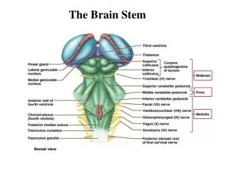
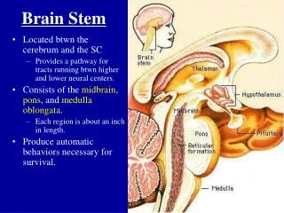
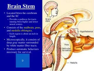
Localization of Brain Stem Lesions. Anatomy of the Brain Stem. Part of the brain that extends from: The rostral plane of the Superior Colliculus To the caudal end of the Medulla Oblongata at the Foramen Magnum Contains Structures: Midbrain Pons Medulla Oblongata.

Localization of Brain Stem Lesions
E N D
Presentation Transcript
Anatomy of the Brain Stem Part of the brain that extends from: The rostral plane of the Superior Colliculus To the caudal end of the Medulla Oblongata at the Foramen Magnum Contains Structures: • Midbrain • Pons • Medulla Oblongata
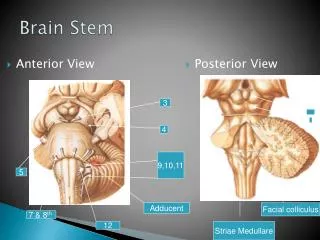
Brain Stem anterior view 1. Optic chiasm2. Optic nerve3. Optic tract4. Medial sulcus of the crus cerebri5. Oculomotor nerve6. Pons 7. Pyramidal eminence of the pons8. Retroolivary fossa9. Oliva10. Posterolateral sulcus11. Decusssation of the pyramids12. Anterolateral sulcus13. Lateral funiculus14. Pyramid15. Foramen caecum16. Middle cerebellar pedunculus17. Trigeminal nerve18. Crus cerebri19. Interpeduncular fossa, • posterior perforate substance20. Mammillary body21. Tuber cinereum22. Infundibulum
Posterior view of the brain stem 1.Pineal gland2.Thalamus ( Pulvinar )3.Superior colliculus4.Inferior colliculus5.Lemniscal trigone6.Frenulum veli7.Superior medullary velum8.Median sulcus9.Gracile tubercle10.Cuneate tubercle11.Posterior intermediate sulcus12.Posteromedian sulcus13.Vagal trigone14.Hypoglossal trigone15.Striae medullares16.Facial colliculus17.Locus coeruleus18.Parabrachial recess19.Crus cerebri20.Inferior collicular brachium21.Medial geniculate body22.Lateral geniculate body23.Suoerior collicular brachium24.Habenula25.Habenular commissure
Brain Stem lateral view 1. Medial geniculate body2. Inferior collicular brachium3. Superior colliculus4. Inferior colliculus5. Superior cerebellar peduncle6. Rhomboid Fossa7. Gracile fascicle8. Cuneate fascicle9. Lateral funiculus10. Pyramid11. Posterolateral sulcus12. Oliva13. Retroolivary fossa14. Bulbopontine sulcus15. Pons16. Trigeminal nerve17. Lateral sulcus of the crus cerebri18. Pontomesencephalic sulcus19. Crus cerebri20. Optic nerve21. Optic tract22. Lateral geniculate body23. Leminiscal trigone24. Middle cerebellar peduncle25. Inferior cerebellar peduncle
Medulla Oblongata (Myelencephalon) • Most caudal Portion of the brainstem • Extends from The Rostral border of the Pons Rostral to the emergence of the first spinal roots Join with the spinal cord at the Foramen Magnum
Vascular supply Barainstems large regional arteries Has three types of branches • Para median branches: supplying midline structures • Short circumferential: supply ventrolateral & lateral surface • Long circumferential: Supply posterior structures & Cerebellum
Brain stem arteries - anterior view 1. Posterior cerebral artery2. Superior cerebellar artery3. Pontine branches of the basilar artery4. Anterior inferior cerebellar artery5. Internal auditory artery6. Vertebral artery7. Posterior inferior cerebellar a.8. Anterior spinal artery • 9. Basilar artery
Para median Bulbar branches (Para median portion) Vertebral artery and Anterior spinal artery Hypoglossal Nucleus Medial longitudinal fascicules The pyramids Inferior Olivary Nucleus (medial part) Lateral bulbar branches (Lateral portion) Intracranial vertebral artery fourth segment or the Posterior inferior Cerebellar artery Occasionally the basilar artery or the anterior Inferior Cerebellar artery
Medullary syndromes Medial Medullary Syndrome Cause:1. Occlusion of ( vertebral a.), (anterior spinal a.), (basilar a. lower segment) 2.Vertebrobasilar dissection 3.Dolichoectasia of the vertebrobasilar system 4. Embolism and meningovascular syphilis
Anterior Spinal a. occlusion (Slide 7) • Ipsilateral pyramid, medial lemniscus, hypoglossal nerve Clinical Picture: • Ipsilateral paresis, atrophy and fibrallation of the tongue the protruded tongue deviates toward the lesion(HN) (away from the hemiplegia • Contra lateral hemiplegia (Py) (face is spared) • Contra lateral loss of position and vibration sense (ML) Pain and temperature spared spinothalamic tract is not affected • Occasional upbeat nystagmus (MLF involvement ) Bilateral involvemnt gives • Quadriparesis • Bilateral LMN lesion of the tongue • Complete loss position and vibration sense
Occasionally: • HN can be spared In Anterior spinal artery occlusion. • Only the pyramids can be damaged givingPure motor hemiplegia • Central facial paresis Corticobulbar fibers descend ipsilaterally before crossing to the facial nucelus of the other side. • Crossed motor hemiparesis Lesions of lower medulla of the crossed fibers of the arm and uncrosseds fibers of to the leg. Lateral Medulllary Syndrome( Wallenberg) Intracranial vertebral artery or posterior inferior cerebellar artery occlusion Causes: • Spontaneous discection of the vertebral artery • Medullary neoplasms Usually metastasis • Cocaine abuse • Abscess • Demyelinating disease • Radionecrosis, Hematoma, trauma, neck manipulations
Characteristic Clinical Picture are: Results of wedge shaped damage to the lateral medulla • Ipsilateral facial hypalgesia & thermoanestesia (Trigeminal spinal n.and tract) Ipsilateral facial pain • Contra lateral trunk & extremity hypalgesia & thermoanesthesial (due to Spinothalmic tract) • Ipsilatral palatal pharyngeal and vocal cord paralysis wit dysphagia and dysarthria (Nucleus Ambiguus) • Ipsilatral Horners syndrome (Descending sympathetic fibers) • Vertigo, nausea, and vomiting (Vestibular nuclei) • Ipsilateral Cerebellar signs (Inferior cerebellar peduncle and cerebellum) • Occasionally Hiccups (Medullary respiratory centers) Diplopia (Lower Pons) Rostral medulla( Severe dysphagia, Hoarsness of voice , Facial paresis) Caudal medulla (Marked vertigo, nystagmus, gait ataxia)09
Rare manifestatios of Wallenberg’s Syndrome: • Wild arm ataxia ( Lateral Cuneate n.) • Ipsilateral limb cllumsiness ( Subolivary area) • Central pain associated with allodynia • Contralateral hyperhydrosis with ipsilatral anhydrosis • Inability to sneeze ( Spinal n.of trigeminal N.) • Loss of taste (N.Tractus Solitarius) lateral zone • Autonomic dysfunction ( N.Tractus Solitarius Medial caudal zone) • Failure of Automatic breating( n. Ambigiuus adjecent Reticular Formation) Ocular motor abnormalities: • Dysfunction of ocular alignment ( Otolithic vestibular n. damage) Elevation of the contralateral eye with out vertical displacement of the ipsilatral eye. Rssulting in diplopia, head tilt , environmental tilt • Torsional nystagmus • Nystagmus • Smooth pursuit and gaze holding abnormality( Cerebe;ar FlloculusParaaflloculusassoing through the inferior peduncle. • Lateropulsion or ipsupulsion • Abnormalities of saccades (Cerebellum –Amplitudes control not speed ) patients have contralateral hypometra and ipsilateral hypermetra
Other lesions • Isolated vertigo with ipsilatral lateropulsion of the trunk (Medial branch of PICA) • Bilateral cerebellar infarction (PICA) Vertigo, Nystagmus Retropullsion,ataxia,upsidedown vision) • Babinski-Nageotte syndrome (Hemimedullary syndrome) L+M syndrome Intracranial vertebral a. • Tegmeental medullary lesion –Medullary satiety • Opalski syndrome LM synd. Ipsilateral hemiplegia Lower med. Lesion f corticospinal tract after pramidal decusation • Lateral pontomedullary syndrome LM synd. + Pontine findigs (Vll +VIII nerves smptoms
THE PONS • Anatomy of the Pons Part of metencephalon Extending caudal plane of striae medullaris posteriorly To pontomedullar sulcus anteriorly Inferrior colliculus dorsally and cerebellar peduncles ventrally Dorsal part referred as Tegmentum Ventral part as Basis pontis or Ponto cerebellar portion Contains Cranial Nerve nuclei,Fiber tracts
Vascular supply Paramedian Vessels 4-6 in number arising from the Basilar a. supply –Medial basal pons, pontine nuclei cortico spinal fibers medial leminiscus Short circumferential a. arise from Basilar a. enter the brachium pontis supply Ventrolateral basis pontis Long circumferential Superior cerebellar a.. Arise from Basilar a. Suply : the dorsolateral pons Brachium pontis Dorsal Retiular formation Periaquidctal region Ventrolateral pontine tegmentum occasionaliy Anterior inferior cerebellara. arise mostly from the basilar a. supply: lateral tegmentum of the lower two thirds of the pons Ventrolateral cerebellum Internal auditory a. arise from Basilar a. Supply: Auditory ,Facial , vestibular Ns
Ventral pontine syndrome (Millard –Gubler syndrome) Lesion of the ventrocaudal pons Involves basis pontis And fascicles of cranial nerves Vll,Vl Contralateral hemiplegia (Pyramidal tract) Ipsiaeral lateral rectus paresis wit diplopia Ipsilateral peripheral facial paresis Raymond syndrome Lesion of the ventromedial pons Affects ipsilaterl Vl N Corticospinal tract Spares Vll N. Ipsilateral rectus paresis Contralateral hemiplegia sparing the face (Pyramidal tract) Pontine Syndromes
Pure Motor Hemiparesis Lacunar infarcts in the basis pontis Involving the corticospinal tract Motor hemiparesis without facial involvement Other lesions that can give similar findings: internal capsule (Po. Limb) Cerebral peduncle Medullary pyramid Vertigo ,dysartira, & gait abnormality favor pontine lesions Dysarthria-Clumsy hand syndrome Vascular leions in the basis pontis At the junction of the upper one third and the lower two thirds Usually lacunar lesions Facial weakness Severe dysarthria Dysphagia Clumsiness and paresis of the hand Similar findings in: Genu of the internal capsule Deep cerebellar hemorhage
Ataxic Hemipresis Lesions basis pontis (U1/3 +L2/3) Lacunar lesions mostly Homolateral ataxia & crural paresis More severe in the lower limb Occasional :Dysarthria, nystagmus, paresthesia Similar findings in: Thalamocapsular lesions Contralat. post.limb. of int. capsule Contralat. Red nucleus Superficial infarcts in the territory of superficial ant.cerebral a. Para central area Locked in syndrome Bilateral ventral pontine lesion Due to: Infarction. Tumor. Trauma. Haemorrhage. Central pontine myelinolysis Quadriplegia Cort.Sp. Lesions bilat. Aphasia involvement of Cort.Bul. Fibers the lower cranial nerve n. Occ. Involvement of Vll N fascicles Patient is fully awake NO damage to the Reticular Formation or supranuclear oculomotoor pathway
Foville sndrome Involves dorsa pontine tegmentum In the caudal third of the pons It consists of: Contralateral hemiplegia due to corticospinal tract invovment Ipsilateral facial palsy Vll N Inabality to move te eye conjugately to ipsilateral side due to Vl N. or paramedian pontine Reticular formation Raymond-Cestan-Chenais syndrome Rostral lesion of the dorsal pons It consists of : Cerbellar signs Ataxia it coarse Rubral tremors Contralatral sensory modalities are reduced ( medial lemniscus & spinothalamic tract) Ventral extension – contralateral hemiparesis (corticospinal tract) Dorsal Pontine Syndrome
Several clinical syndromes exist Unilateral mediobasal infarcts wit Facio-bracio-crual hemiparesis Dysarthria &and homolateral or bilateral ataxia Unilateral mediolatral basal infarcts: ataxia dysarthria slight hemiparesis , ataxic hemiparesis or clumsy hand dysarthria syndrome Unilateral mediocentral or mediotegmental infarcts Clumsy hand –dysarthria syndrome Ataxic hemiparesis Without sensory or eye mov’t disoders hemiparesis with contralateral facial or abducens palsy Bilateral centrobasal infarcts Pseudobulbar palsy & bilateral sensorimotor disturbance Common causes are Small vessel disease, vertebrobasilar large vessel disease & Cardiac embolism less commmonly Paramedian Pontine syndrome
Lateral Pontine syndrome • Marie_Foix Syndrome • Lesions affecting the brachium pontis • Isilatral cerebelar ataxia ( celebellar connections) • Contralatral hemiparesis ( corticospinal tracts) • Contralatral hemianesthesia for pain and tempature ( spinothalamic tracts) Others
The mesencephalon Anatomy of the mesencephalon • Rostrally Superior Colliculus-Mamillary body plane • Caudally the plane just caudal to the Inferior Colliculus • Divided in to: dorsal Tectum the tegmentum and the cerebral peduncle Contains ascending and descending tracts reticular nuclei and well delinated nuclear mases
Vascular supply of the Mecencephalon • Includes Paramedian and Circumferential vessels • Paramedian vessels Arise from the origins of the Posterior Cerebral a. • Thalamoperforating (supplying the thalmus • Pedunclar ( supplying the media peduncle) (Midbrain tegmentum including Oculomotor n. the Red n. & SN) Circumferential a. Circumferential perpendicular aa. • Quadrigemnial aa.(from PCA supply Sup. & Inf. Colliculi) • Superior cerebellar aa. (Supply Cerebral pedunclesBrachium conjunctivum, superior cerebelum) • Posterior chroidal aa. (supply Cereberal Peduncle lat.sup. Colliculi, Thalamus,Choroid Plexus of the third ventricle) • Anterior Choroidal aa.( From Int. Carotid or MCA) Cerebrl peduncle & supramecencephalic structure • Posterior Cerebral aa ( Gives branch to Mecencephalic vesels)
Ventral Cranial Nerve lll Fascicular Syndrome (Weber) Lesion Cerebral Peduncle esp. medial peduncle May damage pyramidal fibers Fascicle of third nerve Consists of: Contralateral Hemiplegia including te lower face(CoS CoB) Ipsilateral oculomotor paresis + parasymp. Cranial N. /// (Dilated pupil) Dorsal Cranial N /// faciclular syndrome(Benedikt) Lesion affecting the tegmentum May affect Brachium conj., Red n. Cranial N. /// Consists of: Ipsilateral oculomotor paredis wit dilated pupil Contralatera Involuntary mov’t like intention temor ,hemichorea, hemiatetosis (Destruction Red n.) Dorsal Red n lesions = Brachium conj. Can give similar findings (Claude synd.) Mesencephalic Syndromes
Dorsal Mesencephalic syndromes Mainly neuroophthalmologic abnormalities (Sylvian aqueduct synd. Parinaud synd.) Commonly seen in: Hydrocephalus Tumors of Pineal origin Consists of : Paralysis of conj. Upward gaze (downward occ.) Pupillary abnormality( usu,Large Convergence retraction Nystagmus o upward gaze Pathalogic lid retractionCollier‘s sign Lid lag “Pseudo abducens palsy” Top of the Basilar Syndrome Oclusive vascular disease rostral BA Usually embolic Giant aneurysms Vasculits Cerbral angiography Gives infarction of: mid brain thalamus portion of temporal and occipital lobe Consists of : Disorders of eye mov’t Pupillary abnormality Behavioral abnormality Visual field defects Motor and sensory deficits