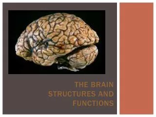
* BRAIN STEM EXTERNAL FEATURES
* BRAIN STEM EXTERNAL FEATURES . Prof. Ahmed Fathalla Ibrahim Professor of Anatomy College of Medicine King Saud University E-mail: ahmedfathala@hotmail.com. OBJECTIVES. At the end of the lecture, students should: List the components of brain stem. Describe the site of brain stem.


* BRAIN STEM EXTERNAL FEATURES
E N D
Presentation Transcript
* BRAIN STEMEXTERNAL FEATURES Prof. Ahmed Fathalla Ibrahim Professor of Anatomy College of Medicine King Saud University E-mail: ahmedfathala@hotmail.com
OBJECTIVES At the end of the lecture, students should: • List the components of brain stem. • Describe the site of brain stem. • Describe the relations between components of brain stem & their relations to cerebellum. • Describe the external features of both ventral & dorsal surfaces of brain stem. • List cranial nerves emerging from brain stem. • Describe the site of emergence of each cranial nerve.
DEVELOPMENT OF BRAIN • The brain develops from the cranial part of neural tube. • The cranial part divides into 3 parts: *FOREBRAIN: subdivides into: 1-Two cerebral hemispheres (cavities: 2 lateral ventricles). 2-Diencephalon (cavity: 3rd ventricle) : thalamus, hypothalamus, epithalamus & subthalamus *MIDBRAIN(cavity: cerebral aqueduct). *HINDBRAIN (cavity: 4th ventricle): subdivides into 1-Pons. 2-Cerebellum. 3- Medulla oblongata.
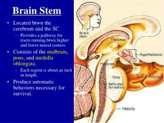
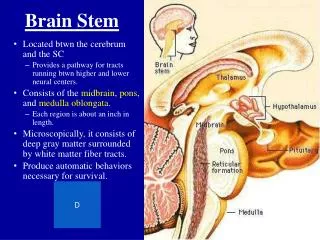
BRAIN STEM • SITE: • It lies on the basilar part of occipital bone (clivus). • PARTS: From above downwards: • Mid brain, pons & medulla oblongata • CONNECTIONS WITH CEREBELLUM: • Each part of brain stem is connected to cerebellum by cerebellar peduncles (superior, middle & inferior).
SAGITTAL SECTION OF BRAIN Thalamus Cerebellum Superior cerebellar peduncle Mid brain Pons Medulla
IMPORTANCE OF BRAIN STEM • Pathway of tracts between cerebral cortex & spinal cord. • Site of origin of nuclei of cranial nerves (from 3rd to 12th). • Site of emergence of cranial nerves (from 3rd to 12th). • Contains groups of nuclei & related fibers known as reticular formation responsible for: control of level of consciousness, perception of pain, regulation of cardiovascular & respiratory systems.
MEDULLA – VENTRAL SURFACE • Ventral median fissure: • It divides the medulla into 2 halves • Its lower part is masked by decussation of most of pyramidal (corticospinal) fibers (75%-90%). • Pyramid: • It lies on either side of ventral median fissure • It is an elevation produced by corticospinal tract.
MEDULLA – VENTRAL SURFACE • Olive: • It lies lateral to the pyramid. • It is an elevation produced by inferior olivary nucleus (important in control of movement). • Nerves emerging from Medulla (4 nerves): • Hypoglossal (12th): between pyramid & olive • Glossopharyngeal (9th), vagus (10th) & cranial part of accessory (11th): dorsolateral to olive (from above downwards) 9 10 12 11
PONS – VENTRAL SURFACE • Basilar sulcus: • It divides the pons into 2 halves. • It is occupied by basilar artery. • Transverse pontine (pontocerebellar) fibers: • It originate from pontine nuclei. • It cross midline & pass through contralateral middle cerebellar peduncle to enter the opposite cerebellar hemisphere.
PONS – VENTRAL SURFACE • Nerves emerging from Pons (4 nerves): • Trigeminal (5th): from the middle of ventrolateral aspect of pons, as 2 roots: a small medial motor root & a large lateral sensory root. • Abducent (6th): at junction between pons & pyramid. • Facial (7th) & vestibulocochlear (8th): at cerebellopontine angle (junction between medulla, pons & cerebellum). Both nerves emerge as 2 roots: from medial to lateral: motor root of 7th , sensory root of 7th , vestibular part of 8th & cochlear part of 8th 5 7 6 8
MID BRAIN – VENTRAL SURFACE • It is formed of a large column of descending fibers (cruscerebri or basis pedunculi), on either side. • The 2 cruracerebri are separated by a depression (interpeduncularfossa). • Nerve emerging from Midbrain (one): • Occulomotor (3rd): from medial aspect of cruscerebri. 3
CLOSED MEDULLA • Cavity: central canal. • Composed of: • Dorsal median sulcus: divdes the closed medulla into 2 halves. • Fasciculus gracilis: on either side of dorsal median sulcus. • Gracile tubercle: an elevation produced at the upper part of fasciculus gracilis, marks the site of gracile nucleus. • Fasciculus cuneatus: on either side of fasciculus gracilis. • Cuneate tubercle: an elevation produced at the upper part of fasciculus cuneatus, marks the site of cuneate nucleus.
OPEN MEDULLA • Cavity: 4th ventricle • On either side, an inverted V-shaped sulcus divides the area into 3 parts (from medial to lateral): • Hypoglossal triangle: overlies hypoglossal nucleus. • Vagal triangle: overlies dorsal vagal nucleus. • Vestibular area: overlies vestibular nuclei.
PONS – DORSAL SURFACE • Separated from the medulla by an imaginary line passing between the caudal margins of middle cerebellar peduncle. • On either side, a sulcus divides the area into 2 parts (from medial to lateral): • Medial eminence: overlies abducent nucleus. • Vestibular area: overlies vestibular nuclei.
MID BRAIN – DORSAL SURFACE • Marked by 4 elevations: • Two superior colliculi: concerned with visual reflexes. • Two inferior colliculi: forms part of auditory pathway. • Nerve emerging from Midbrain (one): • Trochlear (4th): just caudal to inferior colliculus(The only cranial nerve emerging from dorsal surface of brain stem). 4
SUMMARY • The brain stem is composed (from above downwards) of: midbrain, pons & medulla oblongata which are continuous with each other, with diencephalon above & with spinal cord below. • The brain stem is connected with cerebellum through cerebellar peduncles. • The brain stem is the site of cranial nuclei, the pathway of important ascending & descending tracts & the site of emergence of cranial nerves (from 3rd to 12th). • Cranial nerves (with the exception of 4th) emerge from ventral surface of brain stem.
QUESTION 1 • Which one of the following cranial nerves emerges from ventral surface of midbrain? • Occulomotor (3rd). • Trochlear (4th). • Abducent (6th). • Facial (7th).
QUESTION 2 • Regarding the medulla oblongata, which one of the following is correct? • The pyramid is lateral to olive. • The hypoglossal nerve is the most lateral nerve emerging from it. • The cuneate tubercle is lateral to gracile tubercle. • The cerebellum is connected to it by middle cerebellar peduncle.
QUESTION 3 • Which one of the following is the site of the inferior colliculus? • In the ventral surface of medulla, lateral to the olive. • In the dorsal surface of medulla, medial to the vagal triangle. • In the ventral surface of midbrain, lateral to the medial eminence. • In the dorsal surface of midbrain, above the trochlear nerve.