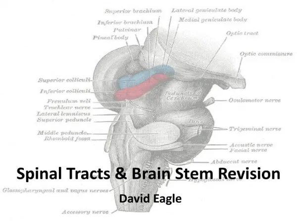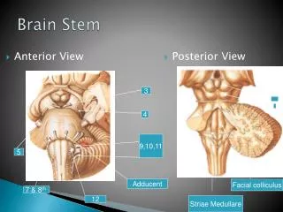
Brain Stem
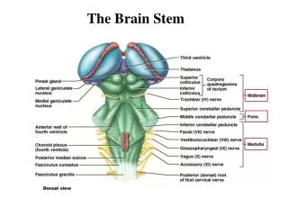
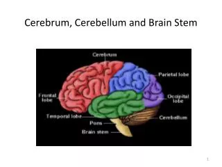
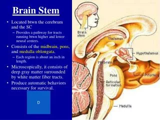
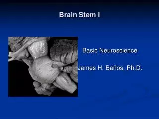
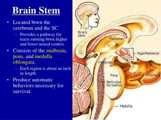
Brain Stem. Located btwn the cerebrum and the SC Provides a pathway for tracts running btwn higher and lower neural centers. Consists of the midbrain , pons , and medulla oblongata . Each region is about an inch in length. Produce automatic behaviors necessary for survival. Limbic System.

Brain Stem
E N D
Presentation Transcript
Brain Stem • Located btwn the cerebrum and the SC • Provides a pathway for tracts running btwn higher and lower neural centers. • Consists of the midbrain, pons, and medullaoblongata. • Each region is about an inch in length. • Produce automatic behaviors necessary for survival.
Limbic System • Includes nuclei and tracts along the border btwn the cerebrum and the diencephalon. • Functions include: • Establishing emotional states • Linking conscious cerebral cortical functions w/ unconscious functions of the brainstem • Facilitating memory storage and retrieval • Limbic system the center of emotion – anger, fear, sexual arousal, pleasure, and sadness.
Protection • What is the major protection for the brain? • There are also 3 connective tissue membranes called the meninges: • Cover and protect the CNS • Protect blood vessels • Contain cerebrospinal fluid • The 3 meninges from superficial to deep: • Dura mater • Arachnoid mater • Pia mater
Skin Galea Aponeurotica Connective Tissue Bone Dura Mater Arachnoid mater
Dura Mater • Tough and leathery. • Most superficial. • 2 layers: • Periosteal attached to the skull • Meningeal true external covering, extends downward and surrounds spinal cord • In several locations, the inner dura mater extends in to the cranial cavity, forming a sheet that dips inward and then returns. These dural folds provide additional support for the brain. Dural sinuses may be found btwn the 2 layers of a dural fold.
Arachnoid and Pia Mater • Arachnoid: • Loose spider-web of connective tissue. • Beneath it is the subarachnoid space – filled with blood vessels and CSF • Pia • Deepest and most delicate • Covers the brain tissue • Follows its every ridge and groove • What do you call an inflammation of the meninges?
Cerebrospinal Fluid • Fills the space btwn the arachnoid and pia mater, as well as the internal cavities of the brain (ventricles) and spinal cord. • Functions: • Shock absorption • Support • Nourishment
Spinal Cord • Functions to transmit messages to and from the brain (white matter) and to serve as a reflex center (gray matter). • Tube of neural tissue continuous w/ the medulla at the base of the brain and extends about 17” to just below the last rib. • Majority of the SC has the diameter of your thumb • Thicker at the neck and end of the cord (cervical and lumbar enlargements) b/c of the large group of nerves connecting these regions of the cord w/ the arms and legs.
Spinal Cord • Notice the gross features of the spinal cord on the right. • 31 pairs of spinal nerves attach to the cord by paired roots and exit from the vertebral canal via the intervertebral foramina.
Cross Sectional Anatomy of the Spinal Cord • Flattened from front to back. • Anterior median fissure and posterior median sulcus partially divide it into left and right halves. • Gray matter is in the core of the cord and surrounded by white matter.
Resembles a butterfly. • 2 lateral gray masses connected by the gray commissure. • Posterior projections are the posterior or dorsal horns. • Anterior projections are the anterior or ventral horns. • In the thoracic and lumbar cord, there also exist lateral horns.
Spinal Meninges Figure 13–3
White Matter • Myelinated nerve fibers. • Allows for communication btwn the brain and spinal cord or btwn different regions of the spinal cord. • White matter on each side of the cord is divided into columns or funiculi. • Typically, they are ascending or descending. • What does that mean?
Spinal Nerves • 31 nerves connecting the spinal cord and various body regions. • 8 paired cervical nerves • 12 paired thoracic nerves • 5 paired lumbar nerves • 5 paired sacral nerves • 1 pair of coccygeal nerves
Spinal Nerves • Each connects to the spinal cord by 2 roots – dorsal and ventral. • Each root forms from a series of rootlets that attach along the whole length of the spinal cord segment. • Ventral roots are motor while dorsal roots are sensory.
Reflex Arcs • A reflex is a rapid, predictable motor response to a stimulus. Unlearned and involuntary. • Example? • Components of a reflex arc: • Receptor site of stimulus • Sensory neuron transmits afferent info to CNS • Integration center 1 or more interneurons • Motor neuron transmits efferent signals to effector • Effector muscle or gland
5 Steps in a Neural Reflex Figure 13–14
Reflexes • Reflexes involving skeletal muscles and somatic motor neurons are somatic. • Reflexes controlled by autonomic neurons are autonomic. • Spinal reflexes are integrated w/i the spinal cord while cranial reflexes are integrated in the brain. • Reflexes may be inborn or learned. • Reflexes may be monosynaptic or polysynaptic. • Difference?
Somatic Reflexes • Let’s look at the muscle spindle reflex and the Golgi tendon reflex and figure out: • What they are? • Why are they somatic? • Are they mono- or polysynaptic? • Are they ipsilateral or contralateral reflexes?
Autonomic Reflexes • May be spinal (e.g., urination and defecation) or modified by higher brain structures. • The thalamus, hypothalamus and brain stem are in charge of multiple reflexes – HR, BP, breathing, eating, osmotic balance, temperature, vomiting, gagging, sneezing. • All are polysynaptic.