Brain stem 1
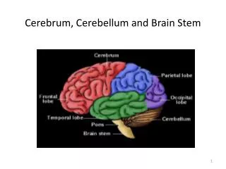
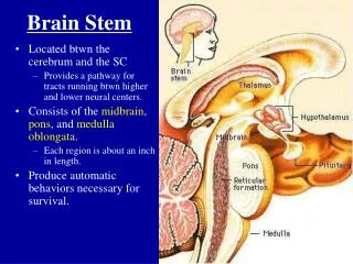
Brain stem 1. Medulla Oblongata. Development of the brain. Brain Stem. Located : between the cerebrum and the SC Consists of : the midbrain, pons, and medulla oblongata . Each region is about an inch in length .

Brain stem 1
E N D
Presentation Transcript
Brain stem 1 Medulla Oblongata
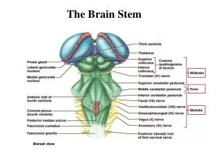
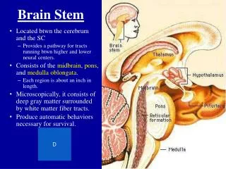
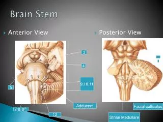
Brain Stem • Located : between the cerebrum and the SC • Consists of :the midbrain, pons, and medulla oblongata. • Each region is about an inch in length.
Microscopically, it consists of deep grey matter surrounded by white matter fiber tracts • CONNECTIONS WITH CEREBELLUM: • Each part of brain stem is connected to cerebellum by cerebellar peduncles (superior, middle & inferior).
FUNCTIONS OF BRAIN STEM • Pathway of tracts between cerebral cortex & spinal cord. • Site of origin of nuclei of cranial nerves (from 3rd to 12th). • Site of emergence of cranial nerves (from 3rd to 12th). • Contains groups of nuclei & related fibers known as reticular formation responsible for: control of level of consciousness, perception of pain, regulation of cardiovascular & respiratory systems.
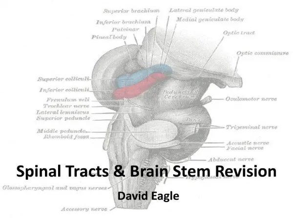
Medulla Oblongata External structure of the medulla • Most inferior region of the brain stem. • Becomes the spinal cord at the level of the foramen magnum. • Medulla is broad above: joins with pons • & narrow below: continuous with spinal cord • Length is about 3cm, width is about 2cm • at its upper end • Surfaces shows series of fissures • Anterior median fissure • Posterior median fissure Medulla oblongata Spinal cord
External features • It is divided into right & left halves by anterior & posterior median fissures • Each half again into ant, post , lat region by anterolateral & posterolateral sulci • The ant. region in the form of a longitudinal elevation called pyramid • Anterior external arcuatefibresrun transversely across upper part of the pyramid • Lateral region shows an oval elevation, the olive. • It is produced by a mass of greymater- inferior olivary nucleus
Pyramid Olive Anterolateral fissure Anterior median fissure
MEDULLA – VENTRAL SURFACE • Ventral median fissure: • Continuation of ventral median fissure of spinal cord • Divides the medulla into 2 halves • Its lower part is masked by decussation of most of pyramidal (corticospinal)fibers (75%-90%). • Pyramid: • An elevation, lies on either side of ventral median fissure • Produced by corticospinal tract.
Olive: • An elevation, lies lateral to the pyramid. • Produced by inferior olivary nucleus (important in control of movement). • Nerves emerging from Medulla (4 nerves): • Hypoglossal (12th): from sulcusbetween pyramid & olive • Glossopharyngeal (9th), vagus (10th) & cranial part of accessory (11th): from sulcusdorsolateral to olive (from above downwards)
MEDULLA – DORSAL SURFACE • The features differ in the caudal part (closed medulla) and the cranial part (open medulla) open medulla closed medulla
CLOSED MEDULLA • Cavity: central canal. • Composed of: 1-Dorsal median sulcus: divides the closed medulla into 2 halves. 2-Fasciculus gracilis: on either side of dorsal median sulcus. 3-Gracile tubercle: an elevation produced at the upper part of fasciculus gracilis, marks the site of gracile nucleus. 4-Fasciculus cuneatus:on either side of fasciculus gracilis. 5-Cuneate tubercle: an elevation produced at the upper part of fasciculus cuneatus, marks the site of cuneate nucleus.
OPEN MEDULLA • Cavity: 4th ventricle • On either side, an inverted V-shaped sulcusdivides the area into 3 parts (from medial to lateral): • Hypoglossal triangle: overlies hypoglossal nucleus. • Vagal triangle: overlies dorsal vagal nucleus. • Vestibular area: overlies vestibular nuclei.
Closed Medulla SubstantiaGelatinosa • Traversed by the Central Canal. • Includes the Motor Decussation. • Includes the Spinal Nucleus of Trigeminal (Trigeminal sensory nucleus) : It is a large sensory nucleus. It is the brain stem continuation of the SubstantiaGelatinosa of spinal cord.
TRIGEMINAL SENSORY NUCLEUS & TRACT • The Nucleus Extends : • Through the whole length of the brain stem and into upper segments of spinal cord. • It lies in all levels of M.O, medial to the spinal tract of the trigeminal. • It receives pain and temperature from face, forehead. • Its tract is present in all levels of M.O. is formed of descending fibers that terminate in the trigeminal nucleus.
PYRAMIDAL DECUSSATION • It is the Motor Decussation. • Formed by pyramidal fibers,(75-90%) cross to the opposite side • They descend in the lateral white column of the spinal cord as the lateral corticospinal tract. • The uncrossed fibers form the ventral corticospinal tract.
MID MEDULLA • Traversed by the Central Canal. • Larger size Gracile & Cuneate nuclei, concerned with proprioceptive deep sensations of the body. • Axons of Gracile & Cuneate nuclei form the internal arcuate fibers; Sensory Decussation. • Pyramidsare prominent ventrally.
SENSORY DECUSSATION • Formed by the crossed internal arcuate fibers • Medial Leminiscus: • Composed of the ascending internal arcuate fibers after their crossing. • Lies adjacent to the middle line ventral to the central canal • Terminates in thalamus. lemniscus = ribbon
Open Medulla • On the ventral aspect : • The pyramid is clear, with medial lemniscus on either sides of middle line dorsal to the pyramid • Inferior Olivary Nucleus: • A convoluted mass of gray matter. Has a hilum directed medially, lies posterolateral to the pyramids & lateral to the medial leminiscus. • It is concerned with the control of movement.
Open Medulla • Its dorsal surface forms: The lower part of the floor of the 4th ventricle. • The Inferior Cerebellar Peduncle is dorsolateral in position, connecting M.O. with cerebellum. • Dorsal and lateral to the Inferior cerebellar peduncle lie the Cochlear nuclei (dorsal and ventral).
Beneath the floor of 4th ventricle lie : 1. Hypoglossal Nucleus lies just lateral to the midline. 2. Dorsal Nucleus of Vaguslateral to the hypoglossal nucleus, contains preganglionic parasympathetic fibers. Medial longitudinal fasciculus liesclose to the midline, ventromedial to the hypoglossal nucleus, dorsal to the medial lemniscus. It links the vestibular nuclei with nuclei of extraocularms. (3,4&6) to help coordination of head & eye movements.
3. Vestibular nuclei complex : is composed of medial, lateral, inferior & superior nuclei, concerned with equilibrium. 4. Nucleus Ambiguus: lie deep to the floor and dorsal to olivary nucleus gives motor fibers to constrictors of the pharynx & intrinsic muscles of the larynx. 5. Solitary nucleus: lie ventrolateral to dorsal nucleus of vagus, receive taste sensation from the tongue along the facial (VII), glossopharyngeal (IX) and vagus (X) cranial nerves.
Cranial Nerves of the Medulla N. solitarious Sensory nucleus for CN VII, IX, X Vestibular nuclei Posterior 1/3 of the tongue Dorsal motor nucleus of X N. ambiguus Motor nucleus for CN IX, X & XI Spinal trigeminal tract CN V, VII, IX, X Stylopharyngeus (lifts pharynx) Sensation behind ear
CN X: Vagus Nerve Dorsal motor nucleus of X Parasympathetic, preganglionic N. solitarious Sensory nucleus for CN VII, IX, X Taste, epiglottis Cardiorespiratory N. ambiguus Motor nucleus for CN IX, X & XI Pharynx Larynx Spinal trigeminal tract CN V, VII, IX, X Ear
Nuclei in the medulla are associated with autonomic control, cranial nerves, and motor/sensory relay. • Autonomic nuclei: 1-Cardiovascular centers Alter the rate and force of cardiac contractions Alter the tone of vascular smooth muscle 2-Respiratory rhythmicity centers Receive input from the pons 3-Additional Centers Emesis, deglutition, coughing, hiccupping, and sneezing