Download
1 / 96
970 likes | 1.19k Vues
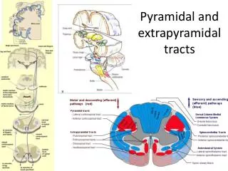
Brain stem: Nuclei and tracts. Medulla. Transitional zone between spinal cord and the medulla. Dorsal expansions of gray matter at the level of pyramids decussation, form the gracile and cuneate nuclei.

E N D
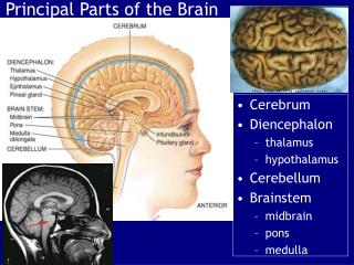
Medulla • Transitional zone between spinal cord and the medulla. • Dorsal expansions of gray matter at the level of pyramids decussation, form the gracile and cuneate nuclei. • Ventral gray horn includes first cervical, spinal root of accessory nerve. Bundles of fibers traversed that pass from the pyramids to the lateral corticospinal (CS) tracts. • The dorsal gray horns of the spinal cord are replaced by spinal trigeminal nuclei. • Above the pyramidal decussation (DP), the medulla has entirely different structures than that in spinal cord.
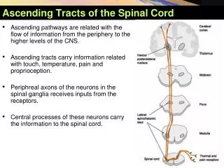
Medial lemniscus system • Gracile fasciculus: touch, proprioception from lower limb (ipsilateral) ends in gracile nucleus. Cuneate fasciculus from upper limb and terminates in cuneate nucleus. There is a point to point projection of the fibers, which served as the anatomical basis for reorganization of the source of a stimulus. • Axons from gracile nucleus and cuneate nucleus form the internal arcuate fibers (IA), cross the midline in the decussation of the medial lemniscus then becomes medial lemniscus. It then runs to the ipsilateral ventral posterior nucleus of thalamus, from there then project to the primary somesthetic cortex of the parietal lobe.
Spinothalamic & Spinotectal Tracts • Spinothalamic tract conveys pain, temperature, from the contralateral side of the body • Spinotectal: same as spinothalamic, conveys somesthetic information to superior colliculus and reticular formation of the midbrain. • The two tracts merge to form spinal lemniscus. Spinal thalamic fibers continue to the ventral posterior nucleus of the thalamus (see previous slides)
Spinoreticular fibers • conveys sensory data from skin and internal organs • It has different route from spinal cord up to thalamus and cerebral cortex. • 1).medial lemniscus system • to ventral posterior thalamic nuclei then somatosensory area of the cerebral cortex. • 2). neospinothalamic system • prominent in mammals, from lamina I, IV-VI (tract cells) in the dorsal horn. It has no collateral fibers to the reticular formation. It ends in ventral posteriolateral division of ventral posterior nucleus of thalamus. • 3). Paleospinothalamic • prominent in all vertibrates. It sends collateral branches to reticular formation, diversed projections.
Spinocerebellar Tracts • Carry proprioceptive signals, dorsal (uncrossed), ventral (crossed). • Dorsal spinocerebellar fibers (L3 above, ) enter the inferior cerebellar peduncle and the ventral spinocerebellar (lumbarsacral region, L-S) continues through pons and enters the cerebellum by superior cerebellar peduncle. Both carry unconscious proprioception to cerebellum (see previous slides).
Accessory (external) cuneate nucleus • Lateral to cuneate nucleus, fibers form these nuclei, cuneocerebellar fibers, enter cerebellum via inferior cerebellar peduncle. Served as supplemental pathway for proprioceptive information from upper limbs.
Inferior olivary complex • Several groups of neurons receive afferent information from different sources and then project them to cerebellum. These neurons are called precerebellar nuclei, which include components of inferior olivary complex. Inferior olivary nucleus is the largest component.
Components • Inferior olivary complex includes • 1). inferior olivary nucleus • 2). medial accessory olivary nucleus • 3). dorsal accessory olivary nucleus
Olivocerebellar fibers • originate from inferior olivary complex, cross in the midline, enter the inferior cerebellar peduncle. Their function is to maintain equilibrium and the stereotyped movements of postural changes and locomotion
DESCENDING TRACTS • Corticospinal tract: • Originate from frontal and parietal lobes, fibers that cross over in the decussation of pyramids (lateral corticospinal tract). Uncrossed fibers (15%, Ventral corticospinal tract), cross at spinal cord level. Major function: motor control
Tracts originate from midbrain • 1). Central tegmental tract: • arises from ipsilateral red nucleus, terminates in inferior olivary complex, relay signals • 2). Axons from contralateral red nucleus (rubrospinal tract) • terminates C2 in human • 3). tectospinal tract: • originates in superior colliculus, cross the midline • 4). tectobulbar fibers: • from superior colliculus, ends in reticular formation of pons and medulla, involves eye movements.
Nuclei of Cranial Nerves • Hypoglossal nucleus • Innervate tongue muscles • Nucleus ambiguus • Of vagus nerve, innervate muscles of soft palate, pharynx, larynx, upper esophagus • Dorsal nucleus • Vagus’ largest parasympathetic nucleus
Dorsal Pons (Tegmentum) • Pons can be divided into: • Ventral (Basal) • Tegmental (dorsal)
Acsending tracts • Medial lemniscus • Twisted when it leaves medullar and enters pons. So the sequence is neck, arm, trunk, and leg from medial to lateral. • Spinal lemniscus • Lateral to the medial lemniscus • Ventral spinocerebellar tract: most lateral, enters cerebellum through the superior cerebellar peduncle
Cerebellar peduncles • Inferior cerebellar peduncle • Inferior cerebellar peduncles enter the cerebellum from the caudal part of the pons.
superior cerebellar • superior cerebellar peduncle and its decussation • Superior cerebellar peduncles (mainly cerebellar efferent fibers) enter the brain stem caudal to the inferior colliculi of the midbrain. The fibers cross the midline in the decussation of the superior cerebellar peduncles. Then most of the fibers continue to the ventral lateral nucleus of the thalamus, from there, project to motor area of the cortex in the frontal lobe. Rest of the fibers end in red nucleus and in the reticular formation. Ascending fibers of superior cerebellar peduncles include ventral spinocerebellar tract as well as fibers from the red nucleus and the mensencephalic trigeminal nucleus.
Nuclei of Cranial Nerves • Trapezoid body from dorsal and ventral cochlear nuclei • Such fibers above end in superior olivary nucleus • Fibers from dorsal cochlear nucleus and superior olivary nucleus form lateral lemniscus
MLF • Medial longitudinal fasciculus • From superior vestibular nucleus, terminates in abducens. Trochlear and oculomotor nerves • Coordinate movements of eyes with the rest of the head
Nuclei of Cranial Nerves • Facial motor nucleus • Abducens nucleus • Spinal Trigeminal Tract and Nucleus • See previous slide