Download
1 / 94
2.28k likes | 9.52k Vues
Ascending Tracts of the Spinal Cord. Ascending pathways are related with the flow of information from the periphery to the higher levels of the CNS. Ascending tracts carry information related with touch, temperature, pain and proprioception.

E N D
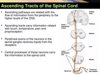
AscendingTracts of theSpinalCord • Ascending pathways are related with the flow of information from the periphery to the higher levels of the CNS. • Ascending tracts carry information related with touch, temperature, pain and proprioception. • Periphreal axons of the neurons in the spinal ganglia receives inputs from the receptors. • Central processes of these neurons carry the information to the spinal cord.
Overview of AscendingTracts Modality:Touch, pain, temperature, proprioception Receptor: Exteroceptor, interoceptor, proprioceptor Primary Neuron: Dorsal root ganglion (spinal ganglion) Secondary Neuron: Spinal cord or brain stem Tertiary Neuron: Thalamus (ventrobasal nuclear complex) Termination:Cerebral cortex, cerebellar cortex, or brainstem
AscendingTracts • Fasciculus gracilis and fasciculus cuneatus • Lateral spinothalamic tract • Anterior spinothalamic tract • Posterior spinocerebellar tract • Anterior spinocerebellar tract • Cuneocerebellar tract • Spinotectal tract • Spinoreticular tract • Spino-olivary tract
FasciculusGracilisandFasciculusCuneatus • Thesetwopathwaysarerelatedwithconsciousproprioception • Proprioception is sense of positionandmovement, sense of vibration, as well as twopointtactilediscrimination.
FasciculusGracilisandFasciculusCuneatus • First order neurons of these pathways are located in the spinal ganglia. • Peripheral processes of these neurons receives information related with position and kinesthesisfrom muscle spindles, golgi tendon organs, joint receptors, and information related with two point discrimination and vibration from receptors such as Pacinian, Meissner, Ruffini corpuscules • The central processes of these axons enter the spinal cord and ascend in the posterior funniculus forming the fasciculus gracilis and fasciculus cuneatus
FasciculusGracilisandFasciculusCuneatus • Axons form the coccygeal, sacral, lumbar and lower six thoracic segments form the fasciculus gracilis, whereas upper six thoracic and cervical segments form the fasciculus cuneatus • Therefore, below T6 there is only fasciculus gracilis; above T6 both fasciculus gracilis (medial) and cuneatus (lateral) exist
FasciculusGracilisandFasciculusCuneatus • Axons of the fasciculus gracilis and cuneatus synapse with the neurons in the nucleus gracilis and nucleus cuneatus • Both nuclei are located at the lower levels of medulla oblangata • Axons of second order neurons from the nucleus gracilis and nucleus cuneatus are called theinternal arcuate fibers which decussate in the caudal medulla to form the medial lemniscus
FasciculusGracilisandFasciculusCuneatus • The medial lemniscus ascends through the pons and midbrain to reach the thalamus. • These axons synapse with the third order neurons at the ventral posterolateral nucleus (VPL)of thalamus. • Axons from VPL traverse the internal capsule and synapse with the neurons in thepostcentral gyrus(somatosensory cortex, Brodmann areas 3,1 and 2)of the cerebral cortex.
FasciculusGracilisandFasciculusCuneatus • Conscious proprioception • 1st order neuron: Dorsal Root Ganglion (Spinal Ganglion) • 2nd order neuron: Nucleus gracilis andcuneatus • Internal Arcuate Fibers decussate and form theLemniscal Decussation • Fibers ascend as the Medial Lemniscus • 3rd order neuron: Thalamus (VPL) • Axons of the third order neurons pass through the Internal Capsule • Termination: Primary Somesthetic Area (Brodmann areas 3, 1, 2)
FasciculusGracilisandFasciculusCuneatus Lesions of these tracts produce the following symptoms: • Posterior funniculus ataxia • Loss of proprioceptive input impairs coordination resulting in ataxia, (walking is clumsy and uncoordinated) • Loss of vibration sense and discriminative tactile sense • Romberg sign • With the eyes closed, patients can not maintain stance as they can not sense the position of the body parts • Lesions of the medial lemniscus produce contralateral signs • Lesions of the posterior funiculus pruduce ipsilateral signs below the level of the lesion
FasciculusGracilisandFasciculusCuneatus • Tabes Dorsalis is a manifestation of syphilis usually affecting the lumbar cord. • Usually there is a degeneration of the dorsal roots and columns leading to a loss of tactile and proprioceptive sensations of the pelvic limbs and ataxia.
LateralSpinothalamicTract • This pathway is related with pain and temperature senses.
LateralSpinothalamicTract • First order neurons of this pathway is located in the spinal ganglia. • Peripheral processes of these neurons receive information related with pain and temperature from the skin and other tissues. • Receptors are free nerve endings. • The central processes of these axons enter the spinal cord and synapse with the neurons in the lamina I, IV and Vof the gray matter. • As entering the tip of posterior horn axons give branches which travel one or two higher and lower segments (at the posterolateral tract of Lissaeur) and synapse at those segments also).
LateralSpinothalamicTract • Axons of second order neurons decussate in the anterior white commissure and ascend in the lateral funiculus as the lateral spinothalamic tract. • These axons synapse with the third order neurons at the ventral posterolateral nucleus (VPL)of thalamus. • Axons from VPL traverse the internal capsule and synapse with the neurons in thepostcentral gyrus(somatosensory cortex, Brodmann areas 3,1 and 2)of the cerebral cortex.
LateralSpinothalamicTract • Somatotopicorganisation:
LateralSpinothalamicTract • Somatotopicorganisation: • In the lateral spinothalamic tract, axons from the sacral sacral segments are most lateral, wheras cervical ones are most medial. • Pain fibers are situated anteriorly and the temperature fibers are posterior.
LateralSpinothalamicTract • Summary of lateral spinothalamic tract: • Pain and temperature • 1st order neuron: Dorsal Root Ganglion (Spinal Ganglion). • 2nd order neuron: Gray matter of the spinal cord (lamina I, IV, V) • Axons of second order neurons decussate at the anterior white commissure. • Fibers ascend as the lateral spinothalamic tract in the lateral funiculus. • 3rd order neuron: Thalamus (VPL) • Axons of the third order neurons pass through the Internal Capsule • Termination: Primary Somesthetic Area (Brodmann areas 3, 1, 2)
LateralSpinothalamicTract • Some fibers of the lateral spinothalamic tract also terminates in other areas of the CNS. • Brainstem reticular formation, cingulate gyrus of the limbic system and insular gyrus • These fibers carry the slow pain impulses. Fast PainSlow Pain Sharp, pricking Dull, burning Group III (A) fiber Group IV (C) fiber Short latency Slower onset Well localized Diffuse Short duration Long duration Less emotional Emotional, autonomic response Not blocked by morphine Blocked by morphine
LateralSpinothalamicTract Clinical note Lesions of this tract produce the following symptoms: • Loss of pain and temperature sense on the contralateral side of the lesion. • The signs are usually detected one or two segments below the level of lesion. • This is due to ascending branches in the posterolateral tract of Lissaeur.
AnteriorSpinothalamicTract • This pathway is related with light (crude) touch and pressure.
AnteriorSpinothalamicTract • First order neurons of this pathway is located in the spinal ganglia. • Peripheral processes of these neurons receives information related withlight touch an pressure. • Receptors are free nerve endings. • The central processes of these axons enter the spinal cord and synapse with the neurons in the lamina I, IV and Vof the gray matter. • As entering the tip of posterior horn axons give branches which travel one or two higher and lower segments (at the posterolateral tract of Lissaeur) and synapse at those segments also.
AnteriorSpinothalamicTract • Axons of second order neurons decussate in the anterior white commissure and ascend in the anterior funiculus as the anterior spinothalamic tract. • These axons synapse with the third order neurons at the ventral posterolateral nucleus (VPL)of thalamus. • Axons from VPL traverse the internal capsule and synapse with the neurons in thepostcentral gyrus(somatosensory cortex, Brodmann areas 3,1 and 2)of the cerebral cortex.
AnteriorSpinothalamicTract • Summary of lateralspinothalamictract: • Lighttouchandpressure • 1storder neuron: Dorsal Root Ganglion (Spinal Ganglion) • 2nd order neuron: Graymatterfothespinalcord (lamina I, IV, V) • Axons of secondorderneuronsdecussate at theanteriorwhitecommissure. • Fibersascend as theanteriorspinothalamictract in theanteriorfuniculus. • 3rd orderneuron: Thalamus (VPL) • Axons of thethirdorderneuronspassthroughtheInternal Capsule • Termination: Primary Somesthetic Area (Brodmannareas 3, 1, 2)
AnteriorSpinothalamicTract Clinical note Lesions of this tract produce the following symptoms: • Loss of light touch and pressure on the contralateral side of the lesion (discriminative touch is preserved) • Patient will not the feel light touch of a piece of cotton or pressure produced in the skin by a blunt object . • The signs are usually detected one or two segments below the level of lesion. • This is due to ascending branches in the posterolateral tract of Lissaeur.
Posterior (Dorsal) SpinocerebellarTract • Modality:Unconscious proprioception • Receptor:Muscle spindle, Golgi tendon organ, joint receptors • 1st Order Neuron:Dorsal root ganglion (Spinal ganglion) • 2nd Order Neuron:Nucleus dorsalis (Clarke’s column) – Located in lamina VII • Ascends ipsilaterally in the posterior part of the lateral funiculus and reaches to the medullla oblangata where it passes through the inferior cerebellar peduncle to reach the cerebellum. • Termination:Cerebellar cortex
Posterior (Dorsal) SpinocerebellarTract • Nucleus dorsalis (Clarke’s column) extends between C8 and L4 segments. • Fibers contributing the posterior spinocerebellar tract below the level of L4 ascends ipsilaterally in the posterior funiculus until they reach L4 and enter the nucleus dorsalis here
Anterior (ventral) SpinocerebellarTract • Modality: Unconscious proprioception • Receptor:Muscle spindle, Golgi tendon organ, joint receptors • 1st Order Neuron: Dorsal root ganglion (Spinal ganglion) • 2nd Order Neuron:Nucleus dorsalis (Clarke’s column) – Located in lamina VII • Majority of the neurons crossat the anterior white commissure but some ascend ipsilaterally. • Fibers reach to the pons where they pass through the superior cerebellar peduncle to reach the cerebellum. • It is accepted that the crossed fibers make a second cross within the cerebellum,therefore, terminating in the ipsilateral part of the cerebellum. • Termination: Cerebellar cortex
CuneocerebellarTract • Modality:Unconscious proprioception from cervical and upper thoracic segments (known as the upper limb equivalent of the posterior spinocerebellar tract) • Receptor:Muscle spindle, Golgi tendon organ, joint receptors • 1st Order Neuron:Dorsal root ganglion (Spinal ganglion) • 2nd Order Neuron:Cuneate nucleus (certain region of the cuneate) • Axons of the second order neurons form the posterior external arcuate fibersand pass ipsilaterally to inferior cerebellar peduncle to reach the cerebellum • Termination:Cerebellar cortex
InSummary • Anterior and posterior spinocerebellar tracts and the cuneocerebellar tract convey impulses related with position and movement to the cerebellum. • This information is used by cerebellum in the coordination of limb movements and maintenance of posture. • These pathways does not reach to the cerebral cortex, and therefore, does not reach counsciousness. • Lesions of these pathways does not produce any disturbance as the conscious pathways cover their function.
SpinotectalTract • Modality: Impulses related with spinovisual reflexes (movement of the eyes and head toward the source of stimulation) – some authors accept that this tract is related with noxious stimuli that results in tissue damage. • Receptor: Various • 1st Order Neuron: Dorsal root ganglion (Spinal ganglion) • 2nd Order Neuron: Spinal cord gray matter (exact laminae are not known) • Axons of the second order neurons cross at the anterior white commissure and ascend anterolateral to the lateral spinothalamic tract. • Termination: Superior colliculus (a visual reflex center located at the level of mesencephalon)
SpinoreticularTract • Modality: Various • Receptor: Muscle spindle, Golgi tendon organ, joint receptors • 1st Order Neuron: Dorsal root ganglion (Spinal ganglion) • 2nd Order Neuron: Spinal cord gray matter (probably at lamina V, VII, VIII of the spinal cord) • Axons of the second order neurons ascend posterior to the lateral spinothalamic tract. • Termination: Neurons of the reticular formation in the medulla oblongata, pons and mesencephalon. • This tract convey various information to the brainstem reticular formation, which plays an important role in influencing levels of consciousness.
Spino-OlivaryTract • Modality:Various • Receptor:Cutaneous receptors as well as muscle spindle, Golgi tendon organ, joint receptors • 1st Order Neuron:Dorsal root ganglion (Spinal ganglion) • 2nd Order Neuron:Spinal cord gray matter (exact lamainae are not known) • Axons of the second order neurons cross at the anterior white commissure and ascend anterior to the lateral spinothalamic tract • Termination:Inferior olivary nuclei in the medulla oblongata – axons of the inferior olivary nuclei cross the midline and project to the cerebellum via the inferior cerebellar peduncle