The Spinal Cord
The Spinal Cord. Basic Neuroscience James H. Baños, Ph.D. Grey and White Matter. Grey and White Matter. Grey Matter = Cell Body. White Matter = Myelinated axon. Grey and White Matter. Grey matter Cortex Nucleus (CNS) Ganglion (PNS) Exception: Basal Ganglia. Grey and White Matter.
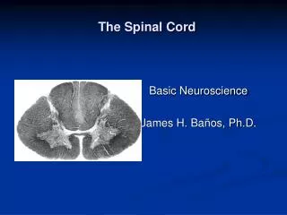

The Spinal Cord
E N D
Presentation Transcript
The Spinal Cord Basic Neuroscience James H. Baños, Ph.D.
Grey and White Matter Grey Matter = Cell Body White Matter = Myelinated axon
Grey and White Matter • Grey matter • Cortex • Nucleus (CNS) • Ganglion (PNS) Exception: Basal Ganglia
Grey and White Matter • White Matter • Nerve (PNS) • Tract (CNS) • Fasciculus/Funiculus -- Group of fibers with common origin and destination • Lemniscus -- Ribbon-like fiber tract • Peduncle -- Massive group of fibers -- usually several tracts
Grey and White Matter • Tracts are named with origin first, then destination • Corticospinal tract -- cortex to spinal cord • Mammilothalamic tract -- Mammilary bodies to thalamus • Spinocerebellar tract -- Spinal cord to cerebellum • Corticobulbar tract -- Cortex to brain stem
General Organization • Spinal cord is SMALL! • 42-45 cm long • 1 CM wide at widest point • Does not extend all the way to the bottom of the spinal column • Pattern of grey/white matter is reversed in the cord • White matter tracts on outside • Grey matter on the inside • Staining reverses this!!!
General Organization White matter (tracts of axons) Grey Matter (cell bodies)
General Organization • Spinal cord is segmented anatomically • Input and output occurs in groups of rootlets arranged in a series longitudinally along the cord • Dorsal rootlets -- Input -- carry sensory information • Ventral rootlets -- Output -- motor neurons
General Organization • Each set of rootlets forms a spinal nerve that innervates a corresponding segment of the body, called a dermatome
General Organization • There are 31 segments in the spinal cord: • 8 cervical (C1 - C8) • 12 Thoracic (T1 - T12) • 5 Lumbar (L1 - L5) • 5 Sacral (S1 - S5) • 1 Coccygeal
General Organization • The spinal cord is housed within the vertebral column
General Organization • Each cord segment has a corresponding vertebra of the same name (e.g., C3) • Spinal nerves enter/exit underneath their corresponding vertebral segment
General Organization • But wait! Something doesn’t add up! • How can spinal nerves exit below their corresponding vertebral segment if the cord is only 42cm-45cm long? • Answer: Spinal nerves extend down to the appropriate vertebral segment forming the cauda equina • This means cord segments and vertebral segments don’t line up
General Organization • Cord is not of uniform thickness throughout its length. Why not?
General Organization • Cord is not of uniform thickness throughout its length. Why not? • Answer: • Segments of the cord innervate parts of the body that differ in complexity • There are fewer white matter tracts lower in the cord.
General Organization Cervical enlargement C5 - T1 Lumbar enlargement L2 - S3
Cord Sections • Segments of the spinal cord have a similar organization, but vary in appearance. • Always know where you are in the cord (i.e., cervical, thoracic, lumbar, sacral)
Cord Sections -- Cervical • Cervical cord is wide, flat, almost oval in appearance. Why?
Cord Sections -- Cervical Enlargement • What’s different about the cervical enlargement . Why? Cervical Cervical Enlargement
Cord Section -- Thoracic • Less White matter than cervical • Rounder appearance • Less prominent ventral horns than cervical enlargement
Cord Section -- Lumbar • Less White matter than thoracic • Rounder appearance • Larger ventral horns, especially in lumbar enlargement Lumbar Lumbar Enlargement
Cord Section -- Sacral • Not much white matter • Mostly grey, although not much of that either
Cross Sectional Organization Posterior intermediate sulcus Posterior median sulcus Tract of Lissauer Anterior white commissure Anterior median fissure
Grey Matter • Laminar • Laminae of Rexed
Grey Matter • Posterior (dorsal) Horn • Intermediate Grey • Anterior (ventral) Horn
Grey Matter: Posterior Horn • Mostly Interneurons • Substantia gelatinosa • Pain/temp proc • Body of the posterior horn • Sensory proc
Grey Matter: Intermediate Grey • Clarke’s Column • T1-L3 • Balance/proprio. • Intermediolateral Column • T1-L3 • Sympathetic neurons
Grey Matter: Anterior Horn • Lower Motor Neurons
White Matter: The “Big Four” Pathways Corticospinal tract Dorsal Columns Spinothalamic tract Spinocerebellar tracts
Corticospinal tract Voluntary motor Dorsal columns/ medial lemniscus Discriminative touch Conscious proprioception Spinocerebellar tract (dorsal and ventral) Unconscious proprioception Spinothalamic tract Pain/temperature The Big Four
Corticospinal Tract Voluntary Motor
Corticospinal Tract • First order neuron (upper motor neuron) originates in precentral gyrus • Passes through internal capsule • 90% decussates in caudal medulla • Lateral corticospinal tract • 10% undecussated • Anterior corticospinal tract • Synapses on second order neuron (lower motor neuron) in ventral gray of the cord • Second order neuron innervates muscle
HAL Motor Homunculus
Motor Homonculus HAL: Arms Legs Head
Corticospinal Tract Spinal Cord Medulla Pons Midbrain
Spinal Cord Medulla Pons Midbrain Corticospinal Tract
Upper & Lower Motor Neurons Motor Ctx • Upper Motor Neuron • Motor Cortex to Ventral Grey Horn • Modulatory influence on stretch reflex arc • Lower Motor Neuron • Ventral Grey Horn to Neuromuscular Junction • Efferent of stretch reflex arc • Helps maintain tone • Sensory Neuron • Stretch receptors in muscle and tendons • Helps maintain tone • Afferent of basic stretch reflex arc UMN Ventral Grey Horn LMN
Upper & Lower Motor Neurons • Maintenance of Tone • Input from stretch receptors causes lower motor neuron to supply tonic stimulation to the muscle • The upper motor neuron modulates this -- will tend to “override” the tonic signal from the sensory neuron UMN LMN
Upper & Lower Motor Neurons • Reflex Arc • Afferent is sensory neuron detecting a sudden stretch • Signal is strong and results in a strong response by the lower motor neuron • Strong signal usually overcomes mild cortical input from the UMN UMN LMN
Upper & Lower Motor Neurons Motor Ctx • Upper Motor Neuron Signs • Spastic paresis • Hypertonia • Hyperreflexia • No muscle atrophy (until perhaps late in the course) • Positive Babinski • Why? • Loss of voluntary UMN signal • Loss of modulation of tone and reflexes by UMN -- the circuit runs unchecked UMN Ventral Grey Horn LMN
Upper & Lower Motor Neurons Motor Ctx • Lower Motor Neuron Signs • Flaccid paresis/paralysis • Muscle fasciculations • Hypotonia • Hyporeflexia • Muscle atrophy • Negative Babinski • Why? • Loss of LMN for voluntary movement • Loss of efferent component of reflex arc and tone pathway UMN Ventral Grey Horn LMN
Babinski’s Sign • In response to stimulation of the sole of the foot, the toes will usually curl downward. • When UMN inhibition is removed, the toes will curl upward (Dorsiflexion). This is referred to as a positive Babinski or presence of Babinski’s sign.
Related Terms… • Spasticity -- Increased muscle tone and increased reflex contraction (UMN) • Clonus -- Rythmic contractions and relaxations seen when a spastic muscle is stretched (UMN)
Basics of Localization • If all limbs are checked for upper and lower motor neuron signs, you can begin to localize lesions • Left-right differences are also very important