Understanding Spinal Cord Tracts: Ascending and Descending Pathways
360 likes | 1.35k Vues
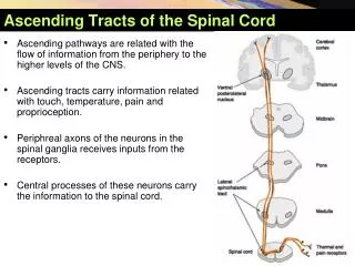
The spinal cord consists of white matter tracts that transmit sensory and motor information via ascending and descending pathways. Ascending tracts carry sensory information from the body to the brain, organized into three neurons: the first order neuron in the spinal ganglia, the second order neuron that ascends through the CNS, and the third order neuron typically in the thalamus, projecting to the cerebral cortex. In contrast, descending tracts convey motor commands from the brain to the spinal cord's lower motor neurons. Key tracts include spinothalamic, corticospinal, and spinocerebellar pathways, which play vital roles in sensory perception and motor control.

Understanding Spinal Cord Tracts: Ascending and Descending Pathways
E N D
Presentation Transcript
Introduction • All white funiculi are composed of longitudinal nerve fibres which are grouped into different functional tracts • For the propose of description they are grouped as ascending and descending tract
Ascending tracts • All modalities of sensations (exteroceptive, propioceptive and interceptive) enters the spinal cord via dorsal root • In spinal cord, they are sorted out and segregated into nerve bundles or tracts in the white mater amd • Ascend from the spinal cord to higher center (brain) • This bundles of ascending fibers are called ascending tract
Contd… • The conduction of information from peripheral sensory ending to higher center consists of 3 neurons • 1st order neuron: has cell body in spinal ganglia. Its peripheral process connects with sensory receptor ending and central process enters the spinal cord via dorsal root and synapses with 2nd order neuron • 2nd order neuron: gives rise to an axon that ascends to a higher level of the CNS, where it synapses with the 3rd order neuron • 3rd order neuron: usually lies in thalamus, gives fibers that passes to the sensory region of cerebral cortex
Contd… • The ascending tracts are • Spinothalamic tract: anterior and posterior/lateral • Posterior white column • Anterior spinocerebellar tract • Posterior spinocerebellar tract • Spino-olivary tract • Spino-tectal tract • Spino-reticular tract
Descending tract Cerebral cortex Corona radiata • The nerve fibers that descend in the white mater from different higher center (medulla, pons, mid-brain and cerebral cortex) are segregated into nerve bundles called the descending tracts • Consists of 2 neuron • Lower motor neuron: situated in the anterior gray column of spinal cord and send axons to innervate skeletal muscles • Upper motor neuron: neurons that convey impulses from higher center to lower motor neuron UMN LMN Decussation muscle
Contd… • Descending tracts are • Pyramidal or corticospinal tract • Extra-pyramidal tract • Rubrospinal tract • Tectospinal tract • Vestibulospinal tract • Olivospinal tract
Spinothalamic tract • 1st order neuron: in dorsal root ganglia • 2nd order neuron: in lamina IV to VII • Axons of these fibers cross the midline • Fibers of lateral spinothalamic tract cross immediately in the same segment • Fibers of anterior spinothalamic tract cross the midline after ascending one or more segment • 3rd order neuron: ventral posterolateral nucleus (VPL) of thalamus • Termination: sensory area of post-central gyrus of cerebral cortex
Contd… • Function • Anterior spinothalamic tract: carry sensation of light/simple touch (non-descriminative/crude) and pressure • Lateral spinothalamic tract: carry sensation of pain and temperature Thalamus Spinothalamic tract
Posterior white column-medial leminiscus pathway • Peculiar that it is formed by the 1st order neurons • 1st order neuron: • axon directly enters the posterior white column of the same side and ascends as fasciulus gracilis (tract of Goll) medially and fasciculus cuneatus (tract of Burdach) laterally • 2nd order neuron: • in the nucleus gracilis and nucleus cuneatus present in the lower part of medulla oblongata • Axon of these neurons cross the midline ascends as medial leminiscus
Contd… • 3rd order neuron: VPL of thalamus • Termination: postcentral gyrus of cerebral cortex • Function: descriminative touch (ability to recognize the two separate points on the skin that are is touched), tactile localization (ability to locate exactly the part touched), stereognosis (ability to recognize the shape of object held in the hand), conscious propriooceptive senses (sense of position and of movement of different parts of body), sense of vibration
Anterior spinocerebellar tract • 1st order neuron: in spinal ganglion • 2nd order neuron: posterior grey column (lamina V and VI) • Most of the axon ascends after crossing the midline • Few fibers do not cross the midline and ascend in the epsilateral side • Termination: cerebellum (cerebellar cortex) through superior cerebellar peduncle • It is believed that those fibers that crossed over to the opposite side in the spinal cord cross back within the cerebellum Anterior spinocerebellar tract
Posterior spinocerebellar tract • 1st order neuron: in spinal ganglion • 2nd order neuron: dorsal nucleus in posterior grey column • fibers do not cross the midline and ascend in the epsilateral side • Termination: cerebellum (cerebellar cortex) through inferior cerebellar peduncle Anterior spinocerebellar tract
Function of spinocerebellar tract • Both spinocerebellar tract convey unconscious proprioceptive sense • Co-ordination and movement of muscles, controlling posture of the body
Spino-olivary tract • 1st order neuron: in spinal ganglion • 2nd order neuron: posterior grey column (lamina IV-VII) • fibers cross the midline • 3rd order neuron: inferior olivary nucleus of medulla • fibers cross the midline • Termination: cerebellum (cerebellar cortex) through inferior cerebellar peduncle • Function: cutaneous and proprioceptive sense Inferior olivary nucleus Spino-olivary tract
Spino-tectal tract • 1st order neuron: in spinal ganglion • 2nd order neuron: posterior grey column (lamina I-V) • fibers cross the midline • Termination: superior colliculus of midbrain • Function: functional significance is un-cleared however is believed to be alternate route of slow pain and also concern with visual spinal reflex (brings movement of head and eye towards the source of stimualtion) Superior colliculus Spino-tectal tract
Pyramidal/Corticospinal tract • Origin (upper motor neuron): motor area of cerebral cortex • Course: • descends successively through corona radiata, internal capsule, midbrain, pons and medulla oblongata
Contd… • At the lower level of medulla • 80-95% cross the midline: lateral corticospinal tract • 5-20% do not cross the midline: anterior corticospinal tract • Termination: interneuron or alpha and gamma neuron of spinal grey mater at various level of spinal cord • Those which do not cross at lower medulla, cross the midline at appropriate spinal level • Function: controls skilful, fine and voluntary movement of opposite half of body
Extra-pyramidal tract • Rubrospinal tract • Origin: red nucleus of midbrain • Fibers immediately cross the midline • Termination: anterior horn cells (alpha and gamma neuron) through interneuron • Function: facilitator to flexor muscles and inhibitory to extensor muscles Red nucleus
Reticulospinal tract • Origin: reticular nuclei of brain stem. Some of them cross and some of them do not cross the midline • Medial reticulospinal tract: reticular nuclei of pons • Lateral reticulospinal tract: reticular nuclei of medulla • Termination: anterior horn cells (alpha and gamma neuron) through interneuron • Function: • Medial: facilitator to extensor and inhibitor to flexor muscles • Lateral: inhibitor to extensor and facilitator to flexor muscles Lateraal recticulospinal tract Medial recticulospinal tract
Tectospinal tract • Origin: superior colliculus of midbrain • Fibers soon cross the midline • Termination: anterior horn cells (alpha and gamma neuron) through interneuron • Function: reflex postural movement (like turning head, moving arm) in response to visual stimulus Superior colliculs eye Tecto-spinal tract
Vestibulospinal tract • Origin: vestibular nuclei (in pons and medulla) • Fibers do not cross • Termination: anterior horn cells (alpha and gamma neuron) • Function: • facilitator to extensor and inhibitor to flexor muscle • Also concerned with the maintenance of equilibrium Deep cerebellar nuclei Vestibular nerve vestibular nuclei Vestibuspinal tract nuclei
Disorder of motor function • Inability to move apart of the body is referred to as paralysis • Monoplegia: paralysis of one limb • Hemiplegia: paralysis of both limb of one side • Diplegia: paralysis of both sides of the body (eg: both arms, or both sides of the face) • Paraplegia: paralysis of both lower limb • Triplegia: paralysis of three limbs • Quadriplegia: paralysis of four limbs • Pathway from cortex to muscle involves at least two neuron; upper motor neuron (UMN) and lower motor neuron (LMN)
LMN paralysis Loss of muscle tone and muscle become flaccid-flaccid paralysis Atrophy of muscle Tendon reflex disappear Fibrillation and fasciculation present UMN paralysis Increased muscle tone and muscle become spastic- spastic paralysis No atrophy of muscle Tendon reflex exaggerated Fibrillation and fasciculation absent LMN paralysis vs UMN paralysis
Syringomyelia • Charaterized by dilation of central canal of spinal cord • Bilateral loss of pain and temperature due to interruption of fibers of spinothalamic tract crossing the midline • But touch, vibratory sense and proprioceptive sense are normal (because ascending tracts in posterior funniculus are unaffected