Ascending tracts
Ascending tracts. Essam Eldin AbdelHady Salama. White Matter in the Spinal Cord. Divided into three funiculi (columns) posterior, lateral, and anterior Each column (funiculus) contains either Ascending (sensory) Descending (motor). White Matter in the Spinal Cord.

Ascending tracts
E N D
Presentation Transcript
Ascending tracts EssamEldinAbdelHadySalama
White Matter in the Spinal Cord Divided into three funiculi (columns) posterior, lateral, and anterior Each column (funiculus) contains either Ascending (sensory) Descending (motor)
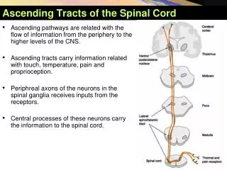
Ascending Tracts • Carry sensory signals up to the spinal cord. • Typically uses 3 neurons • 1st order neuron, detects stimulus and carries it to spinal cord. • 2nd order neuron, continues within spinal cord to the thalamus (the sensory relay station). • 3rd order neuron, carries signal from thalamus to sensory region of cerebral cortex. • Most have names with prefix spino-
Ascending Tracts The first order neurone ,enters the spinal cord through the dorsal root, its cell body lies in the dorsal root ganglion. The central process may make synaptic connections that mediate intersegmental coordination, the main fibres on the ipsilateral side terminate in synaptic contact with the second neurone in the spinal gray matter or in the medulla of brain stem. The second order neurone , its cell body in the spinal cord or the medulla the axons decussate and ascend to the thalamus where terminate upon the third neurone. The third neurone , has its cell body in the thalamus, the axons pass to the somatosensory cortex of cerebral hemisphere.
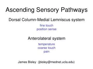
Dorsal column Two tract are recognised Fasciculus gracilis Fasciculus cuneatus They carry, vibration, and proprioception and discriminative touch Receptors are free never ending, muscle spindle , and joint receptors. Cell body of the 1st order neurone lies in the dorsal root ganglion . The central process enters the spinal cord through the dorsal root .
Dorsal column Fasciculusgracilis carries sensation from below T6 Fasciculuscuneatus carries sensation from T6 or higher. They ascend in the spinal cord to relay in; The 2nd order neurone are nucleus gracilis and nucleus cuneatus, Their axons decussate forming the internal arcuate fibers and ascend as the medial lemniscus
Dorsal column The medial lemniscus ascends through the brainstem to relay in ; Ventral posterior (VP) nucleus of thalamus (the 3rd order neuron) then project to the somatosensory cortex of cerebral hemisphere.
Lesion of the posterior column Tables dorsalis Late manifestation of syphilitic CNS Affects lumbosacral dorsal spinal roots and dorsal column of spinal cord. Loss of proprioception leads to unsteady gait (sensory ataxia) exacerbated with closing of eyes. Subacute combined degeneration. Systemic disease due to vitamin B12 deficiency Degeneration of dorsal column causes sensory ataxia Combined with lateral column causes weakness and spasticity of limbs Multiple sclerosis Immune disease affects fasciculus cuneatus of cervical region Leads to loss of proprioception in hands and fingers (asteriognosis)
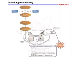
Spinothalamic Pathway Located in the anterior and lateral columns. Carries sensations of pain, temperature, light touch, and pressure. Cell body of the 1st order neurone lies in the dorsal root ganglion . The central process enters the spinal cord through the dorsal root. The 2nd order neuron cell bodies lie in the contralateral dorsal hone. The axons decussate through the ventral white commissure. The axons ascending as spinothalamic tract .
Spinothalamic Pathway In the brain stem it is known as spinal lemniscus, terminate in The ventral posterior nucleus of thalamus as the 3rd order neurone. Finally project to somatosensory cortex.
Spinoreticulothalamic system It is concerned with dull aching pain (slow pain) The sensory impulses from the 2nd neuron ascend and terminate in brain stem reticular formation. Reticulothalamic fibres ascend to intralaminar thalamic nuclei, that in turn activate the cerebral cortex
Spinothalamic tract lesion Syrigomyelia Enlargement of the central canal compressing the adjacent nerve fibres the 2nd order neuron conveying pain and temperature as decussating the ventral white commissure Leads to selective loss of pain and temperature with retaining of light touch and prorioceptive sensation (dissociated sensory loss) with Charcotُs joint . Selective cordotomy or tractotomy is performed to relief intractable pain.
Spinocerebellar tracts ventral &dorsal Carry information from muscle spindle, Golgi tendon organ and tactile receptors to the cerebellum. To control posture and coordination of movement. The dorsal root ganglion is 1st order neuron. The 2nd order neuron cell bodies lie in the base of dorsal horn(Clark ُs) . Terminate directly in the cerebellar cortex (vermis)
Spinocerebellar tracts ventral &dorsal Fibres of dorsal spinocerebellar tractascend ipsilateral, and enter the cerebellum through the inferior cerebellar peduncle. Fibres of ventral spinocerebellar tract , decussate and ascend on contralateral side enter the cerebellum via the superior cerebellar peduncle.
Spinocerebellar tracts lesion Frederic's ataxia Inherited degenerative disease affecting the Spinocerebellar tracts leads to intention tremors ,(involuntary) ,ataxia,
Spino-Olivary Tracts • Project to accessory olivary nuclei and cerebellum. • Contribute to coordination of movement associated primarily with balance.
Spinotectal Tracts • Project to superior colliculi of midbrain. • Involved in reflexive turning of the head and eyes toward a point of cutaneous stimulation.