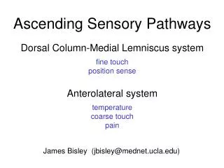
ASCENDING PATHWAYS
ASCENDING PATHWAYS. Ascending Pathways. Three-neuron pathways: Primary sensory neurons: From external receptors Travel through dorsal roots of spinal cord Secondary neurons: Make up tracts in spinal cord and brainstem Tertiary neurons: From thalamus to primary sensory cortex


ASCENDING PATHWAYS
E N D
Presentation Transcript
Ascending Pathways • Three-neuron pathways: Primary sensory neurons: From external receptors Travel through dorsal roots of spinal cord Secondary neurons: Make up tracts in spinal cord and brainstem Tertiary neurons: From thalamus to primary sensory cortex Travel through internal capsule
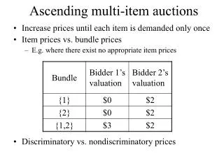
Ascending Pathways • For conscious perception: Spinothalamic system Medial Lemniscal system • For unconscious perception: Spinocerebellar Spino-olivary Spinotectal Spinoreticular
Spinothalamic System • Lateral spinothalamic tract • Anterior spinothalamic tract
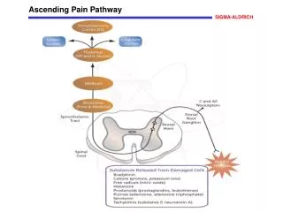
Lateral Spinothalamic Tract • Carries pain and temperature • Primary fibers ascend or descend 1-2 spinal cord segments before synapsing with secondary fibers.
Lateral Spinothalamic Tract • Secondary axons decussate through anterior gray and white commissures. • Secondary axons make up the lateral spinothalamic tract traveling in the lateral column of the spinal cord.
Lateral Spinothalamic Tract • Secondary fibers are joined in brainstem by fibers of the trigeminothalamic tract: (Pain and temperature from face and teeth.)
Lateral Spinothalamic Tract • Secondary fiber collaterals project to reticular formation: Stimulate wakefulness and consciousness. • Secondary fibers project to ventral posterolateral (VPL) nucleus of thalamus.
Lateral Spinothalamic Tract • Secondary fibers synapse with tertiary fibers in VPL. • Tertiary fibers (corticopetal fibers) synapse in postcentral gyrus: Somatic sensory areas 3, 1, 2 • Tertiary fibers form part of internal capsule.
Anterior Spinothalamic Tract • Carries light touch (crude touch), pressure, tickle, itch • Primary neurons may ascend 8-10 spinal cord segments before synapsing with secondary neurons. • Secondary fibers decussate in anterior gray or white commissures.
Anterior Spinothalamic Tract • Secondary fibers ascend to synapse with tertiary fibers in VPL nucleus of thalamus. • Tertiary fibers ascend through internal capsule to primary sensory cortex.
Lateral Spinothalamic Tract • Red • 1° • Blue • 2° • Green • 3°
Lateral Spinothalamic Tract • Red • 1° • Blue • 2° • Green • 3°
Anterior Spinothalamic Tract • Red: • 1° • Blue • 2°
Medial Lemniscus System • Also called posterior column system. • Carries sensations for two-point sensation (fine touch), pressure, and vibration.
Medial Lemniscus System • Primary fibers ascend entire length of spinal cord and synapse with secondary neurons in medulla: Fasciculus gracilis Fasciculus cuneatus
Medial Lemniscus • Red: • Gracilis • Blue • cuneatus
Medial Lemniscus System • Fibers of fasciculus gracilis synapse in nucleus gracilis: Convey sensations from below midthoracic level. • Fibers of fasciculus cuneatus synapse in nucleus cuneatus: Convey sensations from above midthoracic level. Also conveys proprioceptive sensation from arms to cerebellum.
Medial Lemniscus System • Secondary fibers decussate. • Secondary fibers ascend to synapse in VPL of thalamus. • Tertiary fibers ascend through internal capsule to primary sensory cortex.
Posterior Spinocerebellar Tract • Originates in thoracic and upper lumbar regions. • Consists of uncrossed fibers that enter cerebellum through inferior cerebellar peduncles. • Transmits ipsilateral proprioceptive information to cerebellum.
Anterior Spinocerebellar Tract • Originates in lower trunk and lower limbs. • Consists of crossed fibers that recross in pons and enter cerebellum through superior cerebellar peduncles. • Transmits ipsilateral proprioceptive information to cerebellum.
Spino-Olivary Tracts • Project to accessory olivary nuclei and cerebellum. • Contribute to movement coordination associated primarily with balance.
Spinotectal Tracts • Project to superior colliculi of midbrain. • Involved in reflexive turning of the head and eyes toward a point of cutaneous stimulation.
Spinoreticular Tracts • Involved in arousing consciousness in the reticular activating system through cutaneous stimulation.
Stretch (myotactic) Reflex • Muscle spindle = receptor: 3-10 small, specialized intrafusal muscle fibers: Contractile only at ends. Non-contractile center. • Afferent neurons from center of intrafusal fibers travel through dorsal root of spinal nerve to synapse directly with alpha motor neurons of extrafusal fibers in which muscle spindle is embedded.
Stretch (myotactic) Reflex • Afferent neurons from muscle spindle also synapse with ascending fibers within spinal cord. Gamma motor neurons supply intrafusal fibers of muscle spindle: Regulate sensitivity of intrafusal fibers. Gamma neurons are modulated by descending fibers within spinal cord. • Refer to syllabus for specific stretch reflexes.
Golgi-Tendon Reflex • Golgi tendon organs: Encapsulated nerve endings: End with numerous terminal branches with small swellings associated with individual tendon fascicles. Lie within tendons near the muscle-tendon junction. Stimulated when tendon is stretched.
Golgi-Tendon Organs/Reflex • Afferent neurons from Golgi organs pass through dorsal root of spinal nerve and synapse with inhibitory association neurons in posterior gray matter of spinal cord.
Golgi-Tendon Organs/Reflex • Association neurons synapse with alpha motor neurons that innervate muscle fibers associated with tendon. • Causes relaxation of associated muscles and prevents damage to the tendon due to excessive tension.