Transfusion Reactions
Transfusion Reactions. Barbara A. O’Malley, M.D. Associate Director of Transfusion Medicine Harper University Hospital Detroit Medical Center. Complications of transfusion. Acute hemolytic transfusion reaction = rapid destruction of red cells immediately or within 24 hours of transfusion

Transfusion Reactions
E N D
Presentation Transcript
Transfusion Reactions Barbara A. O’Malley, M.D. Associate Director of Transfusion Medicine Harper University Hospital Detroit Medical Center
Complications of transfusion • Acute hemolytic transfusion reaction= rapid destruction of red cells immediately or within 24 hours of transfusion • Most common cause is clerical error: misidentification of the patient • Hypotension, shock, consumptive coagulopathy & acute renal failure • High mortality rate
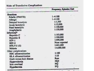
Acute hemolytic transfusion reaction • Rate 1 in 12,000 to 1 in 35,000 reactions per units transfused • Fatal 1 in 100,000 to 1 in 600,000 units transfused • 74% of all fatalities are due to ABO incompatibility • Reported rates may underestimate actual occurrences
Conditions that destroy donor red cells • Naturally occurring antibodies (ABO) • Stimulated alloantibodies (anti-K, Jka) • Autoantibodies • Drug-induced antibodies • Bacterial contamination • Mechanical trauma associated with infusion • Thermal trauma (heat or cold) • Reconstitution of red blood cells with hypotonic solutions • Equipment that damages blood cells extracorporeally
Conditions that destroy recipient red cells • ABO incompatible plasma, cryoprecipitate, or plasma-derived products • (including platelet products which contain plasma) • Infusion of large amounts of hypotonic solutions • Mechanical trauma • (mechanical heart valves, microangiopathic syndromes)
Fever Chills/ rigors Anxiety, feeling of dread Facial flushing Chest pain Abdominal pain Back & flank pain Nausea Vomiting Diarrhea Dyspnea Hypotension Hemoglobinuria Hemoglobinemia Pallor Icterus Oliguria/anuria Pain at transfusion site Diffuse bleeding Jaundice Signs & Symptoms of Acute Hemolytic TR
Management of AHTR • Stop transfusion & maintain venous access • Rapid assessment of pt & requirements for basic & advanced support • Notify transfusion service, collect transfused units & tubing and return to BB • Reconfirm identity of blood units & pt • Collect appropriate patient blood specimens • Supportive approaches
Management of AHTR • Maintain IV fluids at 3000 ml/M2/day with administration of sodium bicarbonate to keep pH >7.0 • Diuretics: • Mannitol (20%) 100ml/ M2 given over 30-60 min, then 30 ml/M2/hr for next 12 hrs. • Furosemide : Adults: 20-80 mg. / Infants & children: 1-2 mg/kg up to an adult dose • Dopamine, low dose: 1-5 mcg/kg/min • Replacement of coagulation factors and platelets • Heparin (controversial in severe DIC) enhances anti-thrombin III
Additional Transfusion Reactions • Delayed hemolytic TR (DHTR) • Febrile nonhemolytic (FNHTR) • Uncomplicated allergic (urticaria) • Anaphylactoid • Anaphylactic (ie. IgA-deficiency) • TRALI (Transfusion-Related Acute Lung Injury) • TACO (Transfusion-Associated Circulatory overload) • Septic Reaction • Post-transfusion (profound thrombocytopenia purpura 1-2 wks post-transfusion, rare) • Iron overload
Delayed Hemolytic TransfusionReactions • Antibody not detected at the time of XM • Rapid secondary boost in antibody level after transfusion: Anamnestic response • 1:2500 to 1:6000 transfusions • Typically cause jaundice at day 5 onwards • May cause hemoglobinuria • Renal failure very rare • Probably under-reported
Febrile Non-Hemolytic TransfusionReaction • Due to cytokines/bioactive proteins in donor plasmaor • Released after WBC antibody in recipient reacts with WBC antigen in product. • Stop transfusion to clinically assess: • Consider acute hemolytic, and bacterial sepsis as part of differential • Report TR to lab, send bag and samples to lab for work up • Treat symptoms with antipyretic (acetaminophen)
Febrile nonhemolytic reactions • Frequent 1: 650 – 1000* • Mainly occur with red cells and platelets • Usually start within 30 minutes • Patient feels cold, shaking, rigors • Temperature rises • Diastolic BP rises • Infected blood should also be considered when this type of reaction occurs *Transfusion 2004; 44: 1-4
Allergic reaction • Frequent 1 in 250 • Usually mild, self-limited • Urticaria Antihistamine prevents • Patient is allergic to something in the donor (foodstuff, medication, protein) or or in the pack (Latex) • May need to use washed products • If Donor is atopic Should not be allowed to donate
Anaphylactic reaction • Severe anaphylaxis • Bronchospasm, shock • 1:20,000 to 1: 50,000 • Usually seen in IgA deficient subjects • They form antibodies to donor IgA • They must receive IgA deficient products
TRALI:Transfusion Related Acute Lung Injury • Severe and potentially fatal reaction to transfusion • Associated with infused granulocyte antibodies and anti-HLA antibodies from donor • Usually donor is multiparous female
TRALI • Chills, fever, dyspnea, non-productive cough, hypotension, 4-6 hours after transfusion • Causes severe respiratory distress and hypoxemia • CXR shows bilateral nodular infiltrates with no cardiac enlargement • Pulmonary wedge pressure is normal
TRALI • Symptoms clear in 24 hrs • CXR clears in 4 days • Estimated frequency 1: 5,000 transfusions • Underdiagnosed, often occurs in patients with other reasons for ARDS and is overlooked
TRALI • Donor antibodies activate Pt’s WBC’s which cause damage to blood vessels in lung tissue • Then fluids and proteins leak into alveolar space/interstitium • Mechanism similar to ARDS
TRALI • Management • Steroids • Aggressive ventilatory support • Hemodynamic support
TRALI • Prevention: • Hemovigilance: Reporting reactions in order to screen involved donor for HLA and neutrophil antibodies • UK: all male donor plasma • ARC: moving in the direction of all male donor plasma
TACO: Transfusion-Associated Circulatory Overload • Cause: • Iatrogenic – physician induced rxn • Fluid(s) administered faster than Pt circulation can accommodate volume load • Some at risk types of pt.’s: congestive heart failure, renal failure, hepatic cirrhosis, normovolemic anemia
TACO • Signs & Symptoms • Cough • Dyspnea • Pulmonary congestion • Headache • Hypertension • Tachycardia • Distended neck veins
TACO • Management: • Place Pt in upright position, if possible, with feet in dependent position • Diuretics • Oxygen • Morphine (if necessary)
TACO • Prevention • Adjust transfusion flow rate based on Pt size and clinical status • Consider dividing unit(s) into smaller aliquot(s) to better space apart blood component(s) pace of transfusion
Septic Reaction • Signs & Symptoms: • Rapid onset of chills & fever • Vomiting, Diarrhea • Profound hypotension, Shock • Cause: • Transfusion of bacterially contaminated blood components • Common problem for platelet concentrates stored at room temp.
Septic Reaction • Management • Obtain blood cultures from Pt • Return blood component bag(s) to blood bank for further laboratory work-up • Treat septicemia with antibiotics • Treat shock with fluids & vasopressors
Septic Reaction • Prevention • Collect, process, store, transport, and transfuse blood components according to contemporary standards of practice (e.g. for FDA standards adhere to cGMP’s – current good manufacturing practices – found in Code of Federal Regulations) • Transfuse blood components within 1 to 2 hrs – do not exceed 4 hrs
Complications of Transfusion • Immunomodulation • Post-operative infections • Transfusion transmitted infections • Graft vs host disease
Transfusion AssociatedGraft Vs Host Disease (TA-GVHD) Symptoms and signs: • Skin rash trunk to extremities day 4 to 30 • Fever day 4 to 23 • Leukopenia day 11 to 30 • Hepatitis • Secondary bacterial / fungal infections • Death day 12 to 65
TA-GVHD • Cause / culprit: transfused lymphocytes • May occur in immunosuppressed or immunocompetent persons • In the immunosuppressed, leukemia and BMT patients are most at risk • In immunocompetent persons: • Donor is a homozygote for HLA haplotype carried by patient
TA-GVHD • In the non-immunosuppressed: • Areas with high rate of HLA homozygosity • Japan ( 1 in 400 ) • Open heart surgery patients • Cases where fresher blood is used • Those receiving blood from close family members (directed donations)
TA-GVHD • Often missed or misdiagnosed • Occurs in patients with other pathology • Preventable by irradiation of cellular blood products: prevents the transfused lymphocytes (Graft) from attacking recipient (Host)
TA-GVHD • Gamma irradiation virtually 100% effective in preventing transfusion-associated GVHD • Irradiate all cellular products transfused to pts at risk • Crosslinks DNA, prevents proliferation of lymphocytes 25Gy to midplane of the blood container, min 15Gy to any point of the irradiated field; max dose not to exceed 50Gy
TA-GVHD • Clear risk • Congenital immunodeficiency • Hodgkin’s disease • CLL treated with fludarabine • Newborns with erythroblastosis fetalis • Directed donations (relatives) • Recipients of HLA matched platelets • Probable risk • Other hematologic malignancies • Solid tumors treated with cytotoxic agents • Genetically homogeneous populations • Premature and possibly full-term neonates • No defined risk • AIDS pts • Immunosuppressive medications
Laboratory Investigation of Transfusion Reactions Sharon Lowry, MT(ASCP)SBB University of Michigan Hospitals October 24, 2008
Why Do a Clerical Check? • Detect labeling errors • Detect patient identification errors • Treat patient for ABO incompatibilities • Prevent companion errors with another patient or another blood unit
Clerical Check - Bedside At the bedside compare • Patient identification • Labels on blood unit
Clerical Check – Blood Bank Compare post-transfusion sample/record Pre-transfusion sample Pre-transfusion test results Blood unit labels Inspect blood unit for color change Confirm IV fluid is saline
Why Do a Visual Check? Hemolysis in patient plasma may be a sign of an acute hemolytic reaction • Antibodies bind to antigens on transfused RBCs • Complement system activated • RBCs are destroyed • Free hemoglobin is released into the plasma Destruction of 5mL of red cells may be visible
Visual Check for Hemolysis • Observe pink or red color inplasma of post-transfusion sample • Compare with pre-transfusion sample
Visual Check Problems Hemolysis observed in plasma may be • Myoglobinemia in trauma • Hemolysis in the donor unit • Underlying condition: AIHA, G6PD • Traumatic draw Collect second sample if hemolysis present
Post ABO Result Compare to pre-transfusion ABO Repeat pre-transfuion ABO if different Explain mixed field agglutination
WBITs Wrong Blood In Tube • Discovered by transfusion reaction • Not discovered if companion sample is same blood type • Missed during pre-transfusion testing when patient has no historical record
CAP TRM.30575 Does the facility have a plan to implement a system to reduce the risk of mistransfusion for non-emergent red cell transfusions? Among the risk reduction options are: Documenting the ABO group of the intended recipient on a second sample collected at a separate phlebotomy Utilizing a mechanical barrier system or an electronic identification verification system that ensures that the patient from whom the pretransfusion specimen was collected is the same patient who is about to be transfused. The use of a second manual banding system, while acceptable, is probably not as effective as the above two options.
Never Events Never should have happened • Incompatible blood transfusions are preventable • Medicare and other insurers will stop paying for added costs of treatment • Patients cannot be charged for error costs
Why Do a DAT? Detect incompatibility • Patient antibodies coat transfused RBCs • Undetected antibodies • Donor antibodies coat patient RBC antigens