Download
1 / 24
240 likes | 336 Vues
Explore the 3D shape variability of healthy and infarcted mouse hearts using Statistical Shape Models. Analyze left ventricle motion, modes of deformation, and results from Tagging MRI. Future research includes improving heart failure detection with advanced techniques.

E N D
3D shape variability of the healthy and infarcted mouse heart Korbeeck, J.M. Eindhoven, July 1st 2004
Contents • Goals; • Anatomy heart; • Modes of LV deformation; • Method: • Tagging MRI; • Statistical Shape Models. • Results; • Future research.
Diseases of the Heart 29% All Other Causes 41% Stroke 7% Cancer 23% Introduction • Infarction major cause of death; • Temporal shape changes mechanical pumping efficiency warning system of heart failure? [From “Stroke Facts 2004: All Americans”, American Heart Association, 2004]
Goals • Study the left ventricle motion of the heart: • Design of Statistical Shape Model algorithm; • Interpretation of shape variability results physiological changes described in literature.
Heart anatomy • Left ventricle (LV) is studied: • Volume corresponds with stroke volume pumping-efficiency; • Blood through entire circulation thickest wall. [From Marieb1997, page 661]
Layers of the heart wall [From Marieb1997, page 658]
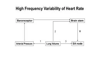
Modes of LV deformation • Deformation: • Radial displacement; • Axial torsion; • Circumferential contraction with long axis extension. • Rotation; • Translation. [From Arts1992]
Tagging MRI (C-SPAMM) • Cine gradient echo MR image of beating heart; • C-SPAMM: • Tag pattern applied by applying magnetic field gradient; • Deformation of the myocardium can be calculated using phase tracking. [From Heijman2004]
Mouse heart • Left ventricle: • Large; • Thick wall. • Right ventricle: • Smaller; • Thinner wall tagging MRI not yet possible. posterior myocardium RV wall RV LV anterior
Statistical Shape Models • Modelling shape and shape variation: • Without shape assumptions. • Shape represented by set of points in time; • Model the variation using PCA.
Principal Component Analysis • Parameterised model: • Reduce dimensionality: • Eigenvectors of covariance; • Eigenvectors main directions; • Eigenvalue variance along eigenvector.
Algorithm • Represent points of 2D image as vector x: • Compute the mean and covariance: • Compute and of S, approximation of x: • Choose t largest eigenvalues such that where fv defines the proportion of total variation
Example [From Cootes2004]
Cardiac Motion Model [From Suinesiaputra2002]
Results • “Normal” (i.e. healthy) heart; • Heart with infarction (LDA occluded by ligation): • Slice through infarction; • Slice above infarction.
Percentage of total Healthy Infarction Above infarction 0.4 0.3 0.2 0.1 0 2 4 6 8 10 Eigenmode Eigenvalues • Healthy heart more eigenvalues mix of more different shape variabilities; • Slice through infarction less deformation modes (mainly translation) caused by infarction; • Great compression component to compensate for infarction.
Eigenmodes healthy Radial compression or compression with long axis extension Translation Rotation or torsion Unknown
Eigenmodes infarction Translation Deviated radial displacement Unknown Unknown
Eigenmodes above infarction Strong compression to compensate for infarction Normal translation Unknown Unknown
Use as filter method Eigenmodes 1-4 • Good approximation with only four eigenmodes ( 95%).
Use as filter method Eigenmode 1 • The end of ventricular systole is almost completely described by the first eigenmode.
Use as filter method Eigenmodes 2-4 • Filtering out of the compression (described by the first eigenmode) works fine.
Future research • Better statistics increment of mice; • Use PCA with foreknowledge; • Analysis of spatial derivatives (circumferential) strain; • 3D tagging MRI/long axis slices; • Link with DTI (fibre tracking).
Future • Better indication of heart failure during a hospital consult after heart dysfunction.