Download
1 / 50
790 likes | 1.77k Vues
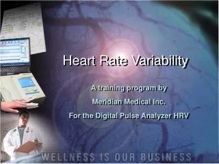
Heart Rate Variability. A training program by Meridian Medical Inc. For the Digital Pulse Analyzer HRV. The Nervous System (Review). The Nervous System is divided into : The Central Nervous System (Brain and Spinal Cord)

E N D
Heart Rate Variability A training program by Meridian Medical Inc. For the Digital Pulse Analyzer HRV
The Nervous System (Review) • The Nervous System is divided into : • The Central Nervous System (Brain and Spinal Cord) • Peripheral Nervous System (divided into the somatic nervous system and the autonomic system)
Nervous System • The spinal cord acts as a communication link between the Brain And the Peripheral Nervous System.
Central Nervous System • Control Centre of the Body • - Relays Messages • - Processes Information • - Compares and Analyzes Information
Peripheral Nervous System • ALL OF THE NERVOUS SYSTEM OUTSIDE THE SPINAL CORD AND BRAIN IS KNOWN AS THE PERIPHERAL NERVOUS SYSTEM • The Peripheral Nervous System consists of neurons NOT included in the brain and spinal cord. • Some Peripheral Neurons collect information from the body and transmit it towards the CNS – Afferent Neurons • Other Peripheral Neurons transmit information away from the CNS – Efferent Neurons
Organs of our body such as the heart, stomach and intestines are regulated by the autonomic nervous system (ANS) ANS is divided into 3 parts : the sympathetic nervous system, the parasympathetic nervous system and the enteric nervous system ANS Review: Autonomic Nervous System
ANS consists of sensory neurons and motor neurons that run between the central nervous system and various internal organs (heart, lungs, viscera, glands) ANS regulates activities that are automatic, or involuntary The nerves of the ANS control functions of the body that are not under conscious control. ANS is concerned with striking a balance or maintaining homeostasis in the functioning of many organs of the body. Autonomic Nervous System
ANS • Most organs controlled by the Autonomic Division are under control of both Sympathetic and Parasympathetic Neurons.
SNS • Often referred to as your 'fight-or-flight' system, your sympathetic nervous system prepares your body for emergencies. • It shunts your blood to your muscles and increases your blood pressure, heart rate and breathing rate, enabling you to cope with stressful situations.
SNS normally functions to produce localized adjustments (such as sweating) and reflex adjustments of the CVS. Under conditions of stress, the entire SNS is activated, producing an immediate, widespread response that has been called the “flight or fight” response. Characterized by the release of large quantities of epinephrine from the adrenal gland, an increase in heart rate, an increase in cardiac output, skeletal muscle vasodilation, cutaneous and GI vasoconstriction, pupillary dilation, and piloerection (hair stands on end) . SNS
Parasympathetic Nervous System • “The Brake” • Main nerves of the PNS are the tenth cranial nerves – the vagus nerves • The PNS returns the body functions to normal after they have been altered by sympathetic stimulation – reverses changes when the danger is over. • The vagus nerves help keep inflammation under control (acetylcholine from the motor neurons suppresses the release of inflammatory cytokines ie: TNF – tissue necrosis factor)
PNS • PNS Stimulation causes: • Slowing down of the heartbeat • Lowering of the blood pressure • Constriction of the pupils • Increased blood flow to the skin and viscera • Peristalsis of the GI Tract
ANS Summary • ANS is a regulatory structure that helps people to adapt to changes in their environment. It adjusts or modifies some functions in response to stress. • ANS helps regulate: • Blood vessels’ size and blood pressure • Hearts electrical activity and ability to contract • Diameter of bronchial tubes and thus air flow in the lungs • Movement and work of the stomach, intestines and salivary glands.
“Autonomic Failure” • What Happens When There Is Autonomic Failure? • Malfunction results from an imbalance between the SNS and PNS divisions. • Aging is associated with several abnormalities in ANS function that can impair the ability to adapt to stress.
Digital Pulse Analyzer • Two Programs • Plethysmograph waveforms to determine arterial elasticity • Heart Rate Variability – Autonomic Nervous System evaluation : Evaluates the level of balance between the branches of the ANS.
What Is Heart Rate Variability? • Heart Rate Variability (HRV) – reflects the heart’s ability to adapt to changing circumstances by detecting and quickly responding to unpredictable stimuli. • Measures the beat-to-beat fluctuations in the rhythm of the heart and the intervals between successive complexes • Sophisticated software enables us to measure imperceptible time differences between normal beats (N-N) • The degree of variance is called HRV.
Recordings should be done at steady-state physiological conditions to produce comparable data Measurements should be done in either a supine or comfortably sitting relaxed position, limiting body movements, conversations and mental activities HRV 5 Minute Analysis
HRV • Heart rate variability (HRV) refers to the beat-to-beat alterations in heart rate. • Under resting conditions, the ECG of healthy individuals exhibits periodic variation in R-R intervals
HRV • HRV is a noninvasive test of cardiovascular autonomic regulation. • Specifically, HRV is a measurement of the interaction between sympathetic (i.e., "fight or flight" energy mobilization) and parasympathetic (i.e., the opposite of the sympathetic activity or "relaxation" response) activity in autonomic functioning
HRV • Low HRV has been shown in numerous longitudinal studies to be related to a higher mortality rate in both healthy and unhealthy subjects. • It can be a predictor of all-cause mortality.
Cardiac disease (Post MI, CHF,LVH) Diabetes Epilepsy Asthma Connective tissue disorder (Lupus) Rheumatoid Arthritis Nutritional deficiencies Genetics Long-term alcoholism Chronic renal disease Hypertension Depression Anxiety/panic attacks Chronic pain Constipation Silent inflammation ANS Disorders displayed in Low Variability
Time Domain / Frequency Domain • According to the Task Force of the European Society of Cardiology and North American Society of Pacing and Electrophysiology in 1996, there are two methods of analysis of HRV data; • Time Domain • Frequency Domain
X X X X RR n-1 RR n RR n+1 ECG X X X X X X X X X X X X X X Time Domain • Normal to Normal (N-N) beats are measured - we capture the QRS complexes resulting from the sinus node depolarization.
TIME DOMAIN MeanHRT (bpm) MeanNN (ms) SDNN (ms) RMS-SD (ms)
Mean HRT • Average Heart Rate over the 5 minute test period. • Reference Range [59.5 – 95.5]
MeanNN (ms) • Measurement in milliseconds the average time between two regular heartbeats. • 628.2ms – 1008 ms (normal range) • <628.2 = seen in tachycardia • >1008 = seen in bradycardia
SDNN – Standard Deviation of Normal to Normal Beats • Most common index of overall HRV • Short-term time domain measures of HRV(5 min) are derived from the differences of successive R-R intervals • Highly correlated and considered to provide good estimates of ANS activity • Reflects our ability to respond quickly, dynamically and effectively to a crisis
Report Parameters Relating to HRV • HRV Graph HRV Histogram HR Distribution
SDNN • Poorly defined in literature • General range: • very poor HRV <30 • poor HRV 30 to 50 • moderate HRV 50 to 100 • optimal HRV >100
How To Improve SDNN • Abstaining from smoking and practicing regular physical activity have been shown to raise the SDNN by 20% • Decrease toxins (food and environment) • Psychotherapy / stress reduction • Supplementation with Omega 3 fatty acids (2g/day of fish oil) showed a significant increase in HRV • Wine
WINE??? • A Stockholm research team studied the effect of alcohol consumption (over 1 year) on 102 women under the age of 75 who had survived a heart attack or surgery of blocked arteries. • HRV was highest in women who drank 5 or more grams of alcohol a day (equivalent to more than half a standard unit) and lowest in those who drank no alcohol at all. • Beer and spirits had little impact on HRV
RMS-SD • The square root of the mean squared differences of successive NN intervals • This measure estimates high frequency variations in heart rate in short-term NN recordings that reflects an estimate of parasympathetic regulation of the heart.
Reflects levels of Sympathetic and Parasympathetic activity and their balance Power in 4 frequency bands analyzed: Total Power Very Low Frequency Low Frequency High Frequency . Frequency Domain Analysis (Spectral Analysis)
Spectral Analysis • Frequency-domain measures pertain to HRV at certain frequency ranges associated with specific physiological processes. • Numerous factors in health and disease have an impact on the amplitude and area of each peak (frequency range) on the HRV spectrum. • Ample evidence that the frequency contributions to HRV are altered in illness states and that the degree of alteration correlates with illness severity.
Frequency Domain Analysis • Also called “Power Spectrum” PSD • Produces low frequency, high frequency, very low frequency and total power (specific fuel tanks!) • Magnitudes in the PSD in certain frequency ranges indicate the relative amount of activity in certain parts of the ANS and the level of stress of the subject.
PSD – Total Power (TP) • Normal Range = 7.2 – 9.1ms2 • Total power = overall “vitality” in the system across all frequencies • Total Power: a short-term estimate of the total power of spectral density in the range of frequencies representing the overall activity of the ANS (autonomic nervous system), however, sympathetic tone is considered as a primary contributor. • Power spectral density (PSD) indicates relative power within each frequency domain derived from the waveform analysis of heart rate interval changes.
Decrease Total Power: Decrease of autonomic nerve activation Equates into decreased ability coping with internal/external stress Increased Total Power: Hyper-stimulated state Total Power
VLF = Very Low Frequency • This measure is not well defined in terms of physiological mechanisms. • With 24 hour recordings, it is considered representing sympathetic tone. • There are some findings in shorter recordings VLF has fair representation of various negative emotions, worries, etc.
Low Frequency (LF) • This frequency band can reflect both sympathetic and parasympathetic activity. • LF decrease = produces symptoms of internal energy loss, fatigue, lethargy. Generally a history of sleep deprivation. (adrenal fatigue?) • .
High Frequency (HF) • This frequency band reflects parasympathetic (vagal) tone and fluctuations caused by respiration known as respiratory sinus arrhythmia. • Parasympathetic tone helps to prevent ventricular arrhythmias by maintaining the electrical stability of the heart. • Decreased HF = result of chronic stress, cardiopulmonary aging, heart disease
LF/HF Ratio • Ratio between the power of LF and HF bands • Normal Ratio: 0.6 to 2.4 • Indicates overall balance between sympathetic and parasympathetic system. • Value greater than 2.4 = Sympathetic Dominance • Value less than 0.6 = Domination of the Parasympathetic system.
Autonomic Balance Diagram (ABD) • Most important part in HRV evaluation • Indicating active status of autonomic nervous system (SNS/PNS) Autonomic nerve , PNS , Chemical sensitivity, High blood pressure, chronic pain, headache, Neurogenic indigestion ) High Frequency 1 2 3 5 Normal Acute stress reaction, Anxiety, Panic disorder, 4 6 Due to decreased SNS activity, Internal energy loss, fatigue (temporary) 7 8 9 Low HF. Illness or pre-illness state, diseases in cardiovascular system Low Frequency ※ Numbers 1,6,8,9 are not applicable
SNS(sympathetic nervous system) & PNS(parasympathetic) Balance - SNS and PNS ratio is normal at around 6:4 - Higher SNS: nervous, anxiety, agitation, excitement, increasing blood pressure, headache, etc. - Higher PNS: depression, sluggishness, lethargy, dizziness, etc.
One of the important issues when measuring HRV is the absence of abnormal heartbeat used in interval detection. Only heartbeats originated in the sino-atrial node can be processed to obtain HRV data. Ectopic beats whether premature ventricular contractions (PVCs) or extrasystolic beats or various movement artifacts will give you a false reading. Remember, HRV measures intervals between N-N (normal to normal beats) PVC Reminders