Stomach and esophagus
730 likes | 1.9k Vues
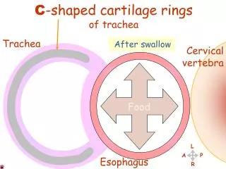
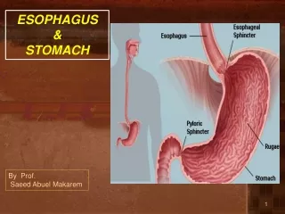
Stomach and esophagus. Esophagus. The esophagus is a tubular structure (muscular, collapsible tube ) about 10 in. (25 cm) long that is continuous above with the laryngeal part of the pharynx opposite the sixth cervical vertebra

Stomach and esophagus
E N D
Presentation Transcript
Esophagus • The esophagus is a tubular structure (muscular, collapsible tube ) about 10 in. (25 cm) long that is continuous above with the laryngeal part of the pharynx opposite the sixth cervical vertebra • The esophagus conducts food from the pharynx into the stomach. Wavelike contractions of the muscular coat, called peristalsis, propel the food onward. • It passes through the diaphragm at the level of the 10th thoracic vertebra to join the stomach • In the neck, the esophagus lies in front of the vertebral column; laterally, it is related to the lobes of the thyroid gland; and anteriorly, it is in contact with the trachea and the recurrent laryngeal nerves • In the thorax, it passes downward and to the left through the superior and then the posterior mediastinum • At the level of the sternal angle, the aortic arch pushes the esophagus over to the midline `
The relations of the thoracic part of the esophagus : • Anteriorly: The trachea and the left recurrent laryngeal nerve; the left principal bronchus, which constricts it; and the pericardium, which separates the esophagus from the left atrium • Posteriorly: The bodies of the thoracic vertebrae; the thoracic duct; the azygos veins; the right posterior intercostal arteries; and, at its lower end, the descending thoracic aorta • Right side: The mediastinal pleura and the terminal part of the azygos vein • Left side: The left subclavian artery, the aortic arch, the thoracic duct, and the mediastinal pleura
Inferiorly to the level of the roots of the lungs, the vagus nerves leave the pulmonary plexus and join with sympathetic nerves to form the esophageal plexus • The left vagus lies anterior to the esophagus and the right vagus lies posterior • At the opening in the diaphragm, the esophagus is accompanied by the two vagi, branches of the left gastric blood vessels, and lymphatic vessels • Fibers from the right crus of the diaphragm pass around the esophagus in the form of a sling. • In the abdomen, the esophagus descends for about 0.5 in. (1.3 cm) and then enters the stomach • It is related to the left lobe of the liver anteriorly and to the left crus of the diaphragm posteriorly.
Blood Supply of the Esophagus • The upper third of the esophagus is supplied by the inferior thyroid artery, • the middle third by branches from the descending thoracic aorta, • and the lower third by branches from the left gastric artery • The veins from the upper third drain into the inferior thyroid veins, from the middle third into the azygos veins, and from the lower third into the left gastric vein, a tributary of the portal vein.
Lymph vessels from the upper third of the esophagus drain into the deep cervical nodes, • from the middle third into the superior and posterior mediastinal nodes, • and from the lower third into nodes along the left gastric blood vessels and the celiac nodes • The esophagus is supplied by parasympathetic and sympathetic efferent and afferent fibers via the vagi and sympathetic trunks • In the lower part of its thoracic course, the esophagus is surrounded by the esophageal nerve plexus.
Gastroesophageal Sphincter • No anatomic sphincter exists at the lower end of the esophagus • However, the circular layer of smooth muscle in this region serves as a physiologic sphincter • As the food descends through the esophagus, relaxation of the muscle at the lower end occurs ahead of the peristaltic wave so that the food enters the stomach • The tonic contraction of this sphincter prevents the stomach contents from regurgitating into the esophagus. • The closure of the sphincter is under vagal control, and this can be augmented by the hormone gastrin and reduced in response to secretin, cholecystokinin, and glucagon.
Stomach - The stomach is a dilated part of the alimentary canal - Between the esophagus and the small intestine
Stomach site It occupies the left upper quadrant mainly in the epigastric region
Shape of stomach • It is roughly J-shaped • Steer horn in obese person • has two openings, the cardiac and pyloric orifices • Two curvatures, the greater and lesser curvatures • Two surfaces, an anterior and a posterior surface
Shape of stomach…..cont Its shape undergoes considerable variation in the same person and depends on - The volume of its contents - The position of the body - The phase of respiration.
Function OF stomach Has three main functions: It stores food (in the adult it has a capacity of about 1500 mL ) It mixes the food with gastric secretions to form a semifluid chyme It controls the rate of delivery of the chyme to the small intestine so that efficient digestion and absorption can take place.
Parts of stomach The stomach is divided into the following parts : 1- Fundus: Dome-shaped Projects upward and to the left of the cardiac orifice It is usually full of gas.
2- Body: Extends from the level of the cardiac orifice to the level of the incisuraangularis (a constant notch in the lower part of the lesser curvature ) 3- Pyloric region divided into: a- Pyloric antrum: - This extends from the incisura angularis to the pylorus
B- Pylorus: - The most tubular part of the stomach - The thick muscular wall is called the pyloric sphincter
Orifices of the stomach - Cardiac orifice - pyloric orifice
Cardiac orifice The cardiac orifice is where the esophagus enters the stomach No anatomic sphincter can be demonstrated here A physiological sphincter physiological mechanism exists that prevents regurgitation of stomach contents into the esophagus
The site of Cardiac orifice • 7th Lt. costal cartilage • 1 inch to Lt. of midline • 45 cm from incisors in the oral cavity. • 10 cm from ant. abdominal wall
pyloric orifice • Present at end of the pyloric canal • On the level of L1 • 1” to the Rt. of the midline. • The circular muscle coat of the stomach is much thicker here and forms the anatomic and physiologic pyloric sphincter • Its position can be recognized by a slight constriction on the surface of the stomach (The pylorus lies on the transpyloric plane).
Pyloric opening…cont The pyloric sphincter controls the outflow of gastric contents into the duodenum. The sphincter receives motor fibers from the sympathetic system and inhibitory fibers from the vagus nerve
Pyloric orifices……cont Function of pyloric opening control by: 1- Hormonal influences from stomach & duodenum 2- Nerve fibers Filling stomach Myenteric fibers relaxation of sphincter
Curvatures of stomach 1- The lesser curvature Forms the right border of the stomach Extends from the cardiac orifice to the pylorus
2- The greater curvature • Much longer than the lesser curvature • Extends from the left of the cardiac orifice, over the dome of the fundus, and along the left border of the stomach to the pylorus
Mucous membrane The mucous membrane of the stomach is thick and vascular and is thrown into numerous folds, or rugae mainly longitudinal in direction The folds flatten out when the stomach is distended.
muscular wall of stomach The muscular wall of the stomach contains longitudinal fibers (outer surface), circular fibers( inner surface), and oblique fibers
Peritoneum of stomach • The peritoneum (visceral peritoneum) completely surrounds the stomach. • It leaves the lesser curvature as the lesser omentum • It leaves the greater curvature as the gastrosplenic ligament and the greater omentum • The gastrosplenic ligament extends from the upper part of the greater curvature to the spleen, and the greater omentum extends from the lower part of the greater curvature to the transverse colon
The lesser curvature is suspended from the liver by the lesser omentum Gastrophrenic ligament between the fundus and the diaphragm.
Relations of stomach Anterior- superior • The anterior abdominal wall • the left costal margin • the left pleura and lung • the diaphragm • the left lobe of the liver
Relations of stomach…cont • Posteriorly = stomach bed • The lesser sac • the Lt. crus of diaphragm • the spleen • the left suprarenal gland • the upper part of the left kidney • the splenic artery • the body of pancreas • the transverse mesocolon • the transverse colon
Blood supply….cont - The arteries are derived from the branches of the celiac artery The celiac trunk arise from the front of the abdominal aorta and its located at the level of T12 to L1 above the pancreas Its 1 cm long
Blood supply for stomach……cont Relations of celiac artery • On each side : celiac ganglia+ lympatic nodes • Crus of diaphragm and lumbar nerves • Its Branches for foregut Main distribution • Lt.gastric.a • Splenic.a • Hepatic.a
Blood supply for stomach……cont 1- The left gastric artery Arises from the celiac artery It passes upward and to the left to reach the esophagus Then descends along the lesser curvature of the stomach It supplies the lower third of the esophagus and the upper right part of the stomach
Blood supply…..cont 2- The right gastric artery • arises from the hepatic artery at the upper border of the pylorus • runs to the left along the lesser curvature. • It supplies the lower right part of the stomach.
Blood supply….cont 3- The short gastric arteries • Arise from the splenic artery (5-7 arteries) • Arises from splenic artery in the gastrosplenic ligament - pass upward in the gastrosplenic to supply the fundus
Blood supply…..cont 4- The left gastroepiploic artery - Arises from the splenic artery before the hilum of the spleen • Passes forward in the gastrosplenic (ligament) • Supply the stomach along the upper part of the greater curvature in the greater omentum 5- The right gastroepiploic artery • arises from the gastroduodenal branch of the hepatic artery • It passes to the left and supplies the stomach along the lower part of the greater curvature in the greater omentum.
Venous drainage The veins drain into the portal circulation The left and right gastric veins drain directly into the portal vein The short gastric veins and the left gastroepiploic veins join the splenic vein The right gastroepiploic vein joins the superior mesenteric vein(which meet the splenic vein behind the neck of pancreas to form the portal vein
Lymphatic drainage • Follow the arteries of stomach • The left and right gastric nodes • The left and right gastroepiploic nodes • The short gastric nodes • All lymph from the stomach eventually passes to the celiac nodes located around the root of the celiac artery on the posterior abdominal wall.
Nerve supply for stomach • The nerve supply includes sympathetic fibers derived from the celiac plexus • parasympathetic fibers from the right and left vagus nerves . • The sympathetic innervation of the stomach carries a proportion of pain sensation • The parasympathetic vagal fibers are secreto-motor to the gastric glands and motor to the muscular wall of the stomach( peristaltic movement) • The pyloric sphincter receives motor fibers from the sympathetic system and inhibitory fibers from the vagus.n.