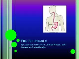
ESOPHAGUS
ESOPHAGUS. MOTILITY DISORDERS. SES LES Peristaltic waves Primar y – deglutition Secundar y – GERD Ter tiary – autonomic muscle control independent of deglutition. MOTILITY DISORDERS. Primary motility disorders Achala s ia : crico pharingian , cardi a Diffuse esophageal spasm

ESOPHAGUS
E N D
Presentation Transcript
MOTILITY DISORDERS • SES LES • Peristaltic waves • Primary – deglutition • Secundary – GERD • Tertiary – autonomic muscle control independent of deglutition
MOTILITY DISORDERS • Primary motility disorders • Achalasia: cricopharingian, cardia • Diffuse esophageal spasm • LES and SES hypertonia • Secondary motility disorders • sclerodermia • diabetes • Parkinson • amiloidosis • colagenosis • miastenia gravis
ACHALASIA • Definition • Lack of LES relaxation + loss of capacity to transmit peristaltic waves, replaced by incoordinated ocntractions
ACHALASIA - pathogeny • Unknown • Degeneration of ganglia cells in Auerbach plexus and vagal motor nuclei. • increased basal pressure in LES and lack of relaxation during deglutition, esophageal pressure and progressive loss of peristalsis • Functional obstruction progressive distension of the esophagus • In time alterations visible on X-Ray exam: • E. dilated, beak-like ending • Level of water = pressure in LES • Long standing = dilated, tortuous
ACHALASIA - pathology • Macroscopic • dilated “botle”, “socket”, initially distal end, followed by all esophagus • Thick wall • Esophagitis due to stasis and fermentationuleration and bleeding • Microscopy • Low or absence of ganglia cells in the nervous plexus of Auerbach
ACHALASIA clinical presentation • 20 – 40y subtle onset • May be asymptomatic • THE ESOPHAGEAL SYNDROME: • Disphagia (intermitent, sometimes very acute, paradoxical!!) • Pain (epigastric, thoracic) - radiates precordial, cervical, ear • Regurgitation (time after eating – depending on dilation of esophagus) – may produce aspiration
ACHALASIA clinical presentation • Periodic development with moment sof “complete” remission • In the stage with competent muscles: fight • predominentpain, disphagia, regurgitation • End stage • Regurgitation • Respiratory: compression, aspiration • Denutrition • Clinical examination: objective findings: nothing
ACHALASIA imagistic Radiology • funcţional • initial • Peristalsis OK • Slow relaxation of cardia • advansed • Upper 1/3 peristalsis • Cabnormal, disorganised contraction that fail to relax cardia • End stage • No peristalsis • Organic • diameter of E • length increased, bent, sinuos • Lower extremity narrow: “beak” or “candle”
Endoscopy • Narrow passage • Does not open, BUT easy passage • cancer
Manometry • No harmonious peristalsis, tertiary waves • High pressure LES • Incomplete relaxation of LES
ACHALASIA – differential dg • Cancer of lower esophagus • Benign peptic stenosis • Difuse esophageal spasmul • Ischemic heart diseases • Respiratory problems • Chagas disease (Trypanosoma cruzi) – damage to myenteric plexus same clinical presentation
Long, unpredictable over 20-30 years 3 stages Dysphagiaand regurgitation Latent clinical stage Megaesophagus Progressive loss of weight : malnutrition Complications general denutrition cachexia regional Mediastinal compression Aspiration: pulmonary complications TB decreased immune reaction: reactivation local Esofagitis UGI bleeding, ulcer Cancer Perforation + mediastinitis ACHALASIA progress
ACHALASIA treatment • Conservative • avoid • Very cold food, very hot food • Rapid eating with large bulky swallows • Decrease LES tonus: nitrates, calcium chanle blockers • Mucosa protection – Sucralfat • Little benefit in time
Forcefull dilation – endoscopic Good initial approach 2 dilations NU surgery RISK: perforation CIND: - long standing disease, tortuous esophagus, association with GERD
ACHALASIA - surgery • Indications • Per primam • Failure after dilation • Extramucosal Heller myotomy • 8-10 cm incision of the muscular wall over the eso-gastric junction • Abdominal/thoracic approach • Fundoplication n(antireflux procedure)
ESOPHAGEAL DIVERTICULA • Classification: • - pharingo-esophageal– Zenker junction between pharinx and esophagus; • - midd-esophageal close to trachea and bronchi; • - epiphrenic (supradiaphragmatic) last 10 cm of esophagus
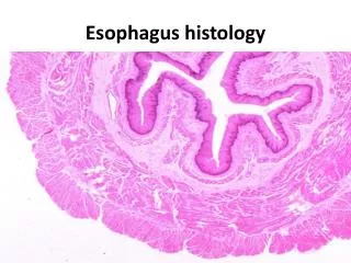
Histology • Structure of the wall: • - true diverticula– all strata of the esophageal wall; • - false (pulsion) – only mucosa and submucosa are present
PATHOGENY • TYPE • - pulsion diverticuladue to high pressure in the lumen + motility disorder • - traction diverticula– inflammatory processes in the vicinity with traction on the wall during scaring.
ZENKER diverticula • fals diverticula • Pulsion type • Weak area: posterior aspect of the pharynx between the inferior constrictir and transversal situatated cricopharingeal = triangle of triunghiul Killian(only mucosa and some fibrotic tissue)
ZENKER diverticula • CLINICAL ASPECT • Initial • Burning sensation, non-productive cough, sensation of foreign body in the neck • Late – big diverticula • Disphagia • Regurgitation • Bead smell • Compression
DIVERTICULUL ZENKER • IMAGISTIC • X-Ray • Endoscopy
ZENKER DIVERTICULA • Complications • Hemorhage • Perforation • Carcinoma • Chronic pulmonary infections
ZENKER DIVERTICULA • TREATMENT • Small • Nothing much • Big, symptomatic, complications • Resection of the diverticula± miotomy of SES • Endoscopic treatment
MIDDLE ESOPHAGEAL DIVERTICULA • Traction type • Middle thoracic • Adjacent inflammatory pathology • ETHIOLOGY • infections • Mediastinal TB (lymph nodes) • Pleural infections • Pericarditis • congenital
PATHOLOGY • Traction type • True diverticula • Lateral wall of the esophagus in the lateral wall • Large mouth to communicate with the esophageal lumen = no retention • Can also be pulsion type (not usual)
IMAGISTIC DIAGNOSTIC • Clinical • asymptomatic • disphagia • Complications • Hemorrhage • Perforation • Cancer • Imagistic • X-Ray • Endoscopy
TREATMENT • Conservative (no clinical signs) • Surgery • Excision • Open or thoracoscopic
EPIPHRENIC DIVERTICULA • Inferior esophagus • PATHOGENY • Associated with motility disorders – achalasia, difuse esophageal spasm • Combines: high pressure + lack of relaxation
EPIPHRENIC DIVERTICULA • DIAGNOSTIC • Similar to thoracic type • +manometry for motility disorder • TREATMENT • Rezection + myotomy • +tratament of associated disease
Physiologic reflux Normal in some instances, but quickly cleared More often standing and while awake LES tonus influenced by different factors: GERD
GERD • Diagnostic: • Presence of symptoms • Endoscopic demonstration of esophagitis • LES
GERD • PATHOGENY • A. Mechanic failure of LES – valve effect • Inadequate LES pressure • Inadequate length of LES • Abnormal position of cardia • Brahiesophagus • B. Inefficient clearance – 4 factors: • Gravity • Normal peristaltic movements of the esophagus • Salivary gland production • Positioning of distal esophagus in abdomen
GERD PATHOGENY • C. Gastric reservoir • Gastric distention: • Decreases the length of LES • Causes: chewing gum, low saliva production (Sjogren), motility disorders. • High Gastric pressure • Outlet syndrome – pyloric stenosis, vagotomy • Diabetic gastroparesis • Prolonged gastric stasis • Miogenic causes (diabetes, neuromuscular problesm, anticholinergic medication, etc) • Non-miogenic causes (vagotomy, antropyloric disfunctin, duodenal motility disorders, duodeno-gastric reflux) • Gastric hypersecretion – exposure to low pH
GERD: E mucosa aggression • Gastric juice: • Low pH – long term exposure • Pepsine – proteolytic at pH<2 • Duodenal reflux: • Billiary salts – E not used to deal with high pH • Pancreatic enzymes • Differences • HCl, billiary acidsmucosal permeability • pepsine, tripsinemucosal erosions • Very little correlation between symptoms and endoscopic appearance of lesions.
GERD complications • ESOPHAGUS: prolonged exposure • esofagitis • strictures • Barrett • RESPIRATORY: repeated aspiration pneumonia, pulmonary fibrosis • Pathological seen lesion correlate with • 1. LES pressure (sphincter volume) • 2. Acid + bile is more aggressive toward mucosa
GERD symptoms • Digestive • Retrosternal burning pain pirozisul • Regurgitations (according to body position) • Dysphagia (edema, stenosis, damaged peristalsis) • Respiratory symptoms • Chronic cogh • Senzation of lack of air • Horse voice (chronic laryngitis) • Wheezing • Unusual symptoms • Nausea, vomiting • Full stomach • Atypical thoracic pain
GERD imagistic • Barium meal- esophagus, stomach and duodenum • Reflux of barium • Hiatus hernia • Strictures • Associated lesions
GERD endoscopy • Every patient with dysphagia - compulsory • Esophagitis • gr I – congestion of mucosa, no ulcerations • gr II – linear ulcerations bordered by granulation tissue that bleeds on touch • gr III – confluent ulcerations with isles of normal mucosa • gr IV - stenosis
GERD – Barrett esophagus • Epitelial metapasis: • Normal squamos cell epithelium – into gastric columnar epithelium • Biopsy: metaplasia, displasia, adenocarcinoma
GERD and hiatus hernia • HH often associated with GERD (main symptom) • Sliding • Rolling – not often associated with reflux (cardia normal) • Combined
GERD - manometry • Stationary manometry • Evaluation of LES • pressure<6mmHg • Total length in abdomen < 1cm • Total LES <2cm • Sphincter area volume • Primary motility disorders (achalasia, difuse spasm) • GERD induced motility disorders RGEafect SEI/ peristaltica E/ amplitudinea contracţiilor
GERD manometry • 24 hours ambulatory manometry • Better diagnostic of motor dysfunctin • In non-obstructive dysphagia: non coordinated muscle function • Scintigrapic evaluation of esophageal transit • 10 ml water with Tc99 • Non specific • Quantification of esophageal transit time • Prolonged time in achalasia, sclerodermia, difuse spasm, nutcracker syndrome
GERD pH monitoring • Quantification time with pH<4 • Total time of pH<4 exposure • Frequency of exposure • Duration of epsiodes >5 minutes • Longest period of reflux • Association with events • Acid relfux: pH<4 • Alkaline reflux pH>7 • Pletismography: quantification of billiary reflux
GERD differential diagnstic • Achalasia –dysphagia + lack of esophageal empting (Rx + endoscopy) • Esophageal cancer: dysphagia (endoscopy) • Hiatus hernia: may be clinical silent • Esophageal diverticula motility disorders, regurgitation (Rx, endoscopy) • UGI pathology • Ischemic heart disease • Pneumonia and other respiratory problems
GERD treatment • Medical – first step (no evaluation) • Minor changes in habits • Raised position in bed • Avoid very tight clothing • Small frequent meals • Dine before 6pm and small quantity • Loose weight • Avoid alcohol, smoking, coffeee, tea, pepermint, chocolate, fat • Protective tratement for mucosa (alginat – creates a barrier)+ antacides (may relieve symptoms but rebound) • Promotilic medication
REFLUXUL GASTROESOFAGIAN • Step II persistent symptoms) • Endoscopy +/- Rx studies for complications; manometry • PPI – decrease gastric acid output (high recurrence when stop) – continuous medication with periods without. Long term: increases risk of hyperplastic gastric polyps • Persistent symptoms: aggressive exploration • TREATMENT: aggressive • Medical • Surgical
GERD surgery • Indications • Persistence of endoscopic lesions in spite of aggressive medication • Young patients – long term treatment • No response to treatment • FUNDOPLICATION