Download
1 / 18
560 likes | 5.17k Vues
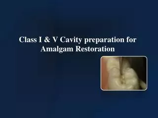
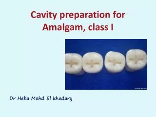
Cavity preparation for Amalgam, class I. Dr Heba Mohd El khodary. Definition.

E N D
Cavity preparation for Amalgam, class I Dr HebaMohd El khodary
Definition Cavity preparation is defined as the mechanical alteration of a defective, injured or disease tooth in order to best receive a restorative material which will reestablish a healthy state of the tooth including esthetic corrections where indicated, along with normal form and function.
What are class I lesions? • Caries in occlusal pits and fissures of posterior teeth • Caries in the occlusaltwo-thirds of the facial and lingual of posterior teeth • Caries in the lingual pits of maxillary incisors
Diagnosing Class I Caries -Softeningat the base of a pit or fissure (WHO). -Opacitysurrounding the pit or fissure, enamel appears chalky when dried. -Softened enamel that may be flaked away by explorer. -Brown-gray enamel ( caused by lateral spread of caries into dentin ). -Radiographic evidence.
INCIPIENT CLASS I CAVITY • Small carious lesion in the central fossa of primary molars with all other teeth being sound. • May be made without local anesthetic. • A No. 329 or No. 330 bur is used. • Restored with amalgam or a resin modified glass ionomer. • A preventive resin restoration may be done if needed.
PIT OR FISSURE CLASS I CAVITY Outline form • should include all fissures, areas of caries, pits and developmental grooves and should be dovetailed. • Facial & lingual margins should not extend more than halfway between central groove and cusp tips(one- quarter to one- third of the intercuspal width) • Leave marginal ridges supported by dentin. • Keep bur perpendicular to the occlusal plane of the tooth while preparing the outline
The extension of the occlusal portion of the cavity preparation depends on the primary molar involved: • The occlusal portion usually is extended about one half the way across on the primary maxillary and mandibularfirst molar.
For the primary mandibular second molar, extend the step completely across the occlucal surface.
-The primary maxillary second molar preparation includes only the nearest occlucal pit. The oblique ridge is not included unless undermined with carious lesions.
Resistance form • Proper form prevents fracture of tooth and restoration during function. • Slightly rounded line angles 2. • Enamel supported by sound dentin. • Weak tooth structure is removed. • Amalgam requires a thickness of at least 1.5 mm to prevent fracture of the material (0.5 mm beneath dentino enamel junction 1). • The walls converge slightly with the greater width at the pulpal floor 3.
Retention Form • Correct retention form ensures that the restoration will not dislodge due to lifting or tipping forces. • Facial and lingual walls are parallel or slightly convergent.
Finish of the enamel walls The cavosurface angle 4 for amalgam restoration should be 90-110 degrees. This is called a butt joint. What is the cavosurface angle 4? (The angle formed by the junction of a cavity wall and the surface of a tooth).
Convenience Form • The cavity allows operator to visualize all remaining caries and to use the proper instruments in the preparation when condensing the amalgam.
DEEP-SEATED CLASS I CAVITY • Plane back the enamel that overhangs the extensive lesion. • The cavity should extend through out the remaininggrooves and anatomical occlusal defects. • The carious dentine should next be removed with large round burs or spoon excavators. • Then , the cavity walls should be finished as previously described. • With deep carious lesions and near pulp exposures, the depth should be covered with a biocompatible base material for adequate thermal protection of the pulp.
Lab Guidelines for Class I • Mark pits and fissures with a sharp pencil Using a 329 or 330 bur drop to length of bur in a pit. • Follow fissures at depth of bur. • Place dove tails mesial and distal. • Diverge mesial and distal walls. • Take prep to 1.5 mm depth. • Smooth walls and floor.