Download
1 / 58
580 likes | 621 Vues
Learn about the plasma membrane's role as a barrier and its selective permeability, membrane models, fluidity, protein functions, and more in cell biology.

E N D
Cytology Membrane Structure and Function
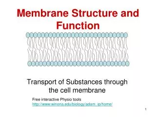
Overview: Life at the Edge • The plasma membrane • Is the boundary that separates the living cell from its nonliving surroundings
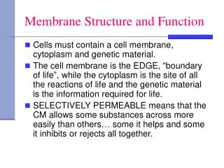
The plasma membrane exhibits selective permeability • It allows some substances to cross it more easily than others
Membranes have certain properties: • They act as a barrier between the cell and its environment, allowing a complex organized system to exist inside the cell. • They permit the passage of selected substances into and out of the cell. • They flex, bend and flow to allow the cell to change shape.
Membranes have certain properties • They act as a barrier between the cell and its environment, allowing a complex organized system to exist inside the cell. • They permit the passage of selected substances into and out of the cell —selective permeability. • They flex, bend and flow to allow the cell to change shape.
Membrane Models: Scientific Inquiry • Membranes have been chemically analyzed • And found to be composed of proteins and lipids
Phospholipids • Are the most abundant lipid in the plasma membrane • contain both hydrophobic and hydrophilic regions
WATER Hydrophilic head Hydrophobic tail WATER Figure 7.2 • Scientists studying the plasma membrane • Reasoned that it must be a phospholipid bilayer
Hydrophobic region of protein Phospholipid bilayer Figure 7.3 Hydrophobic region of protein • In 1972, Singer and Nicolson • Proposed that membrane proteins are dispersed and individually inserted (in a mosaic pattern) into the phospholipid bilayer
The fluid mosaic model of membrane structure • States that a membrane is a fluid structure with a “mosaic” of various proteins embedded in it
Freeze-fracture studies of the plasma membrane • Supported the fluid mosaic model of membrane structure
Freeze-fracturing show particles embedded in Membrane Further reading: http://www.hillstrath.on.ca/moffatt/bio3a/cellbio/phase1.htm 2020/1/4
Lateral movement (~107 times per second) Flip-flop (~ once per month) (a) Movement of phospholipids Figure 7.5 A The Fluidity of Membranes • Phospholipids in the plasma membrane • Can move within the bilayer
Membrane proteins EXPERIMENT Researchers labeled the plasma membrane proteins of a mouse cell and a human cell with two different markers and fused the cells. Using a microscope, they observed the markers on the hybrid cell. RESULTS Mouse cell Mixed proteins after 1 hour Human cell Hybrid cell + CONCLUSION The mixing of the mouse and human membrane proteins indicates that at least some membrane proteins move sideways within the plane of the plasma membrane. Figure 7.6 • Proteins in the plasma membrane • Can drift within the bilayer
Membrane Proteins and Their Functions • A membrane • Is a collage of different proteins embedded in the fluid matrix of the lipid bilayer
Myelin sheath membrane (metabolically inactive) shows little particles (protein) embedded 2020/1/4
Transport. (left) A protein that spans the membrane may provide a hydrophilic channel across the membrane that is selective for a particular solute. (right) Other transport proteins shuttle a substance from one side to the other by changing shape. Some of these proteins hydrolyze ATP as an energy source to actively pump substances across the membrane. (a) ATP (b) Enzymatic activity. A protein built into the membrane may be an enzyme with its active site exposed to substances in the adjacent solution. In some cases, several enzymes in a membrane are organized as a team that carries out sequential steps of a metabolic pathway. Enzymes Signal transduction. A membrane protein may have a binding site with a specific shape that fits the shape of a chemical messenger, such as a hormone. The external messenger (signal) may cause a conformational change in the protein (receptor) that relays the message to the inside of the cell. (c) Signal Receptor Figure 7.9 • An overview of four major functions of membrane proteins
(d) Cell-cell recognition. Some glyco-proteins serve as identification tags that are specifically recognized by other cells. Glycoprotein
Membrane carbohydrates • Interact with the surface molecules of other cells, facilitating cell-cell recognition
1 Transmembrane glycoproteins Secretory protein Glycolipid 2 Golgi apparatus Vesicle 3 Plasma membrane: Cytoplasmic face 4 Extracellular face Transmembrane glycoprotein Secreted protein Membrane glycolipid Figure 7.10 • Membrane proteins and lipids • Are synthesized in the ER and Golgi apparatus ER
Membrane structure results in selective permeability • A cell must exchange materials with its surroundings, a process controlled by the plasma membrane Body cells Blood capillary Red blood cells
The Permeability of the Lipid Bilayer • Hydrophobic molecules • Are lipid soluble and can pass through the membrane rapidly • Polar molecules • Do not cross the membrane rapidly
Transport Proteins • Transport proteins (channel or carrier) • Allow passage of hydrophilic substances across the membrane
Concept 7.3: Passive transport is diffusion of a substance across a membrane with no energy investment
(a) Diffusion of one solute. The membrane has pores large enough for molecules of dye to pass through. Random movement of dye molecules will cause some to pass through the pores; this will happen more often on the side with more molecules. The dye diffuses from where it is more concentrated to where it is less concentrated (called diffusing down a concentration gradient). This leads to a dynamic equilibrium: The solute molecules continue to cross the membrane, but at equal rates in both directions. Molecules of dye Membrane (cross section) Equilibrium Net diffusion Net diffusion Figure 7.11 A • Diffusion • Is the tendency for molecules of any substance to spread out evenly into the available space
(b) Diffusion of two solutes. Solutions of two different dyes are separated by a membrane that is permeable to both. Each dye diffuses down its own concen- tration gradient. There will be a net diffusion of the purple dye toward the left, even though the total solute concentration was initially greater on the left side. Equilibrium Net diffusion Net diffusion Net diffusion Equilibrium Net diffusion Figure 7.11 B • Substances diffuse down their concentration gradient, the difference in concentration of a substance from one area to another
Osmosis is the diffusion of water through a selectively permeable membrane. Water tends to diffuse from a dilute region (higher water potential) to a more concentrated region (lower water potential).
Water Balance of Cells Without Walls • Tonicity /concentration strength (tonicity) of a solution: • Is the ability of a solution to cause a cell to gain or lose water • Has a great impact on cells without walls
If a solution is isotonic • The concentration of solutes is the same as it is inside the cell • There will be no net movement of water • If a solution is hypertonic • The concentration of solutes is greater than it is inside the cell • The cell will lose water • If a solution is hypotonic • The concentration of solutes is less than it is inside the cell • The cell will gain water
Hypotonic solution Hypertonic solution Isotonic solution (a) Animal cell. An animal cell fares best in an isotonic environ- ment unless it has special adaptations to offset the osmotic uptake or loss of water. H2O H2O H2O H2O Normal Shrunk / Shriveled Lysed/ burst Figure 7.13 • Water balance in cells without walls https://www.youtube.com/watch?v=Y_w07A7chnk
Animals and other organisms without rigid cell walls living in hypertonic or hypotonic environments • Must have special adaptations for osmoregulation Paramecium has contractile vacuoles to remove excess water
Water Balance of Cells with Walls • Cell walls • Help maintain water balance
(b) Plant cell. Plant cells are turgid (firm) and generally healthiest in a hypotonic environ- ment, where the uptake of water is eventually balanced by the elastic wall pushing back on the cell. H2O H2O H2O H2O Turgid(normal) Flaccid Plasmolyzed Figure 7.13 • Water balance in cells with walls
If a plant cell is turgid • It is in a hypotonic environment • It is very firm, a healthy state in most plants
If a plant cell is flaccid • It is in an isotonic or hypertonic environment
Facilitated Diffusion: Passive Transport Aided by Proteins • In facilitated diffusion • Transport proteins speed the movement of molecules across the plasma membrane
EXTRACELLULAR FLUID Channelprotein Solute CYTOPLASM (a) A channel protein (purple) has a channel through which water molecules or a specific solute can pass. Figure 7.15 • Channel proteins • Provide corridors that allow a specific molecule or ion to cross the membrane
Solute Carrier protein (b) A carrier protein alternates between two conformations, moving a solute across the membrane as the shape of the protein changes. The protein can transport the solute in either direction, with the net movement being down the concentration gradient of the solute. Figure 7.15 • Carrier proteins • Undergo a subtle change in shape that translocates the solute-binding site across the membrane
Concept 7.4: Active transport uses energy to move solutes against their gradients
The Need for Energy in Active Transport • Active transport • Moves substances against their concentration gradient • Requires energy, usually in the form of ATP
The sodium-potassium pump • Is one type of active transport system
Passive transport. Substances diffuse spontaneously down their concentration gradients, crossing a membrane with no expenditure of energy by the cell. The rate of diffusion can be greatly increased by transport proteins in the membrane. Active transport. Some transport proteins act as pumps, moving substances across a membrane against their concentration gradients. Energy for this work is usually supplied by ATP. ATP Diffusion. Hydrophobic molecules and (at a slow rate) very small uncharged polar molecules can diffuse through the lipid bilayer. Facilitated diffusion. Many hydrophilic substances diffuse through membranes with the assistance of transport proteins, either channel or carrier proteins. • Review: Passive and active transport compared Figure 7.17
Maintenance of Membrane Potential by Ion Pumps • Membrane potential • Is the voltage difference across a membrane
– EXTRACELLULAR FLUID + – ATP + H+ H+ Proton pump H+ + – H+ H+ + – CYTOPLASM + H+ + – • An ion pump that can generate a electrical potential • Is a transport protein that generates the voltage across a membrane Figure 7.18
Cotransport: Coupled Transport by a Membrane Protein • Cotransport • Occurs when active transport of a specific solute indirectly drives the active transport of another solute