Download
1 / 67
890 likes | 1.01k Vues
Liver diseases in pregnancy. By Mahmoud Gaber Ass. Lecturer of internal medicine. Introduction. Severe liver disease in pregnancy is rare. Up to 3% of all pregnancies are complicated by liver disorders.

E N D
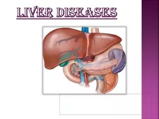
Liver diseases in pregnancy By Mahmoud Gaber Ass. Lecturer of internal medicine
Introduction • Severe liver disease in pregnancy is rare. • Up to 3% of all pregnancies are complicated by liver disorders. • Pregnancy-related liver disease is the most frequent cause of liver dysfunction in pregnancy and provides a real threat to fetal and maternal survival.
Normal physiological changes in pregnancy • Size and appearance of liver unchanged • Blood flow to liver unchanged despite increased maternal blood flow. • Lab.: Alkaline phosphatase increases 1.5 – 4 times above normal. No change in bilirubin. AST/ALT unchanged. Decreased serum albumin.
Normal physiological changes in pregnancy (cont.) • Altered protein synthesis PT unchanged Decreased antithrombin III, protien S & increased fibrinogen prothrombotic state Decreased ceruloplasmin Decreased transferrin Increased cholesterol production (HDL, VLDL, LDL, total cholesterol, triglycerides) • Drug metabolism is unpredictable.
By physical examination : Spider angiomata 60-70% Palmar erythema (63% white, 39% black women) due to hyperestrogenaemia Liver is not palpable Jaundice is NOT normal
Classification of liver diseases in pregnancy • Pregnancy-related liver diseases • Hyperemesis gravidarum • Intrahepatic cholestasis of pregnancy(ICP) • Pre-eclampsia and eclampsia • HELLP syndrome • Acute fatty liver of pregnancy(AFLP)
Pregnancy-unrelated liver diseases Pre-existing liver diseases • Cirrhosis and portal hypertension • Hepatitis B and C • Autoimmune liver disease • Wilson’s disease Liver diseases co-incident with pregnancy • Viral hepatitis • Biliary disease • Budd-Chiari syndrome
Timing of liver diseases related to pregnancy 1st trimester 2nd trimester 3rd trimester Hyperemesis graviderum Intrahepatic cholestasis of pregnancy preeclmpsia HELLPsyndrome Acute fatty liver of prgnancy
Hyperemesis Gravidarum • Defined as intractable vomiting in the first trimester of pregnancy resulting in dehydration, ketosis, and weight loss of 5% or more. • occurs in 0.3–2 % of all pregnancies usually within the first trimester. • The cause remains unclear but abnormal gastric motility, hormonal factors, and changes in the autonomic nervous system are all probably involved. • Liver involvement is seen in about 50%-60% of patients with HG .
Risk factors • increased body-mass index (BMI), • psychiatric illness, • molar pregnancy, • pre-existing diabetes, • multiple pregnancies. • Hyperthyroidism is seen in about 60% of cases of hyperemesis gravidarum (↑ HCG )
Prognosis • No long-term sequelae of liver dysfunction have been described in patients with pregnancies complicated by HG and liver disease.
Biochemical abnormalities: • AST & ALT can be raised upto 20 times the upper limit of normal, usually < 1000 IU. • Raised serum urea and creatinine • Hypokalaemia. • metabolic alkalosis • These resolve on resolution of vomiting. • Imaging studies of the liver in patients with HG are usually unremarkable. • Persistent symptoms beyond week 18 should warrant consideration of a gastroscopy to exclude mechanical obstruction.
Complications 1. Dehydration 2. Malnutrition 3. Mallory-weiss tear of oesophagus 4. Thromboembolic disorders 5. Peripheral neuropathy 6. Hypoglycaemia 7. Acute renal failure 8. Wernicke’s encephalopathy (vit B1 deficiency) 9. Pontine myelinolysis(rapid correction of hyponatraemia)
Management of HG • ttt is supportive and includes intravenous rehydration, antiemetics (Promethazine[C], metoclopramide [B], ondansetron[B] ) and gradual reintroduction of oral intake. • Vitamin supplementation, especially thiamine, is mandatory to prevent Wernicke’s encephalopathy. • No benefit in outcomes is seen with the use of steroids. • If progressive weight loss, jaundice, or persistent tachycardia occurs despite treatment, termination of the pregnancy should be considered.
Intrahepatic cholestasis of pregnancy • Defined as cholestatic disorder characterized by pruritus, elevated serum aminotransferases and bile acid levels with onset in the second or third trimester of pregnancy, and spontaneous relief of signs and symptoms within two to three weeks after delivery. • The incidence is upto 1.5% of pregnancies.
Aetiology • ICP is associated with abnormal biliary transport across the canalicular membrane, the etiology of which is probably heterogeneous • Hormonal factor female sex hormones induce cholestasis and inhibit the bile salt export pump. • Genetic factor the multidrug resistance protein 3 (MDR3) mutation has been identified in 15% of Cases. • Exogenous factor use of progesteron
Clinical presentation • Pruritus is the primary clinical symptom of ICP with a predilection for the palms of the hands and soles of the feet, and is not associated with any specific skin lesions. • Jaundice typically develops 1 – 4 weeks after the onset of pruritus. • Steatorrhea may be seen along with fat malabsorption, which may lead to vitamin K deficiency resulting in a prolonged prothrombin time and postpartum hemorrhage. • Abdominal pain, malaise and other constitutional symptoms are uncommon. • The incidence of gallstone formation and cholecystitis is higher in women with a history of ICP than in the normal population .
The importance of this disorder is related to its effects on the fetus (placental insufficiency, premature labor and sudden fetal death). • Maternal prognosis is good and symptoms resolve rapidly after delivery, accompanied by normalization of serum liver tests .
Biochemical abnormalities: • AST & ALT can be increased by 2-10 fold above the normal level. • Total bile acid levels may increase 10–100 times above the normal range (< 10 μmol/L). • Gamma glutamyl transpeptidase is normal or modestly elevated in half of the patients. • Serum alkaline phosphatase may rise up to 7–10 times above normal. • Ultrasound reveals no biliary duct dilation and hepatic parenchyma appear normal
Management • Therapy is directed at alleviating pruritus in the mother. • Many pharmacological agents have been used in the treatment of ICP. These include phenobarbital, cholestyramine [C] (8-16 g/d) and dexamethasone (12 mg 4 times daily for 7 days followed by a tapering dose). All these agents showed some limited clinical benefit but also had significant side effects. Vitamin K deficiency was observed with the use of cholestyramine in high doses
Management (cont.) • Ursodeoxycholic acid (UDCA) [B] has been used successfully in the treatment of cholestasis . • UDCA has been shown to improve impaired hepatocellular secretion with a standard 15-mg/kg/day dosage. A larger dosage, 20 to 25 mg/kg/day has been shown to be effective with no adverse affects on either mother or baby.
Management (cont.) • Delivery has been recommended in the 37th to 38th week in ICP. • But the UK Guideline for Obstetric Cholestasis 2006 stated that "there are insufficient data to support or refute the popular practice of 'early' (37 weeks of gestation) induction of labour aimed at reducing late stillbirth". The timing and risks of delivery should, therefore, be discussed on an individual basis.
Pre eclampsia and eclampsia : • Pre-eclampsia is a multisystem disorder characterised by hypertension and proteinuria (greater than 300 mg in 24 h) after 20 weeks of gestation and/or within 48 h after delivery. Oedema is no longer needed for the diagnosis of pre-eclampsia. • Presence of seizures differentiates eclampsia from preeclampsia. • Liver involvement occurs in 20–30% of patients and always indicates severe preeclampsia with significant perinatal morbidity and mortality.
Pre eclampsia and eclampsiaClinical features • Hypertention & oedema • Right upper abdominal pain, • Headache, • Nausea, and vomiting.
Pre eclampsia and eclampsia Investigations • Aminotransferase could be upto 10 times the upper limit of normal • Bilirubin concentrations are rarely increased. • Liver biopsy is not indicated, which shows characteristic microscopic changes involve periportal areas with sinusoidal fibrin thrombi, haemorrhage, and hepatocellular necrosis.Portal thrombosis and haemorrhage can also be present. • Liver biochemical profile usually normalises within 2 weeks of delivery.
Pre eclampsia and eclampsia Complications • Maternal hypertensive crises • Renal dysfunction, • HELLP syndrome, • Subcapsular haemorrhage, hepatic rupture or infarction, • Seizures • Increased perinatal morbidity and mortality.
Pre eclampsia and eclampsia Management • No specific therapy is needed for the hepatic involvement. • Its only significance is as an indicator of severe disease with need for immediate delivery to avoid eclampsia. • The only effective treatment for preeclampsia is delivery of the fetus and placenta. However, if mild preeclampsia is evident before fetal lung maturity at 36 wk gestation, one may consider expectant management in ICU. • Pharmacological agents used in preeclampsia include antihypertensives such as calcium channel blockers. • Magnesium sulfate may be administered if eclampsiadevelops
HELLP Syndromepathogenesis • The initial source of the insult is unknown, but all patients have evidence of endothelial injury with fibrin deposit that causes a microangiopathic hemolytic anemia and platelet activation and consumption, leading to thrombocytopenia. • The pathogenesis of liver involvement in HELLP syndrome is unknown. A complex chain of events is initiated in the liver by intravascular fibrin deposition with sinusoidal obstruction, associated with hypovolemia, which is demonstrated by a decrease in the liver blood flow on doppler examination in patients with preeclampsia, who have subsequently developed HELLP syndrome . • Hepatic ischemia causes hepatic infarction, subcapsular hematomas and intraparenchymatous hemorrhage, which may result in hepatic rupture.
HELLP SyndromeDiagnosis • Haemolysis • Microangiopathic haemolytic anaemia (CBC Presence of fragmented (schizocytes) or contracted red cells with spicula in the peripheral blood smear ) • ↑ Indirect bilirubin o LDH > 600 U/l. • Haptoglobin < 25 mg/dl. • Elevated liver enzymesAST > 70 U/l. • Platelets < 100,000/mm3 • The prothrombin time or INR remains normal unless there is evidence of DIC or severe liver injury. • A positive D-dimer test in the presence of preeclampsia has recently been reported to be predictive of patients who will develop HELLP syndrome.
HELLP SyndromeClassification Two classifications for the HELLP syndrome are commonly used The Tennessee System 1 is based on the assessment of the following parameters: AST>70 U/L, LDH >600 U/L, platelets <100,000/ mm3. • There are two forms: complete (all elements present) partial (one or two elements present). Mississippi system1 AST > 40 U/L and LDH > 600 U/L and: • Class I: platelets < 50 x 109/L • Class II: platelets 50 – 100 x109/L • Class III: platelets 100 – 150 x109/L. 1-Lancet 2010; 375: 594–605
HELLP SyndromeManagement • Delivery is the only definitive therapy. • At greater than 34 weeks’ gestation or if there is any evidence of multiorgan dysfunction, DIC, renal failure, abruptio placentae or fetal distress, there is consensus that immediate delivery should be done. • at less than 34 weeks gestation, Controversy exists. Generally, if a reassuring fetal and maternal status exists, delivery may be delayed for a course of steroid. • Platelet transfusion ?? indicated when the platelet count is less than 20,000/mm3, if less than 50,000/mm3 and cesarean delivery necessary, or with any significant bleeding.
HELLP SyndromeManagement(cont.) • Steroid therapy ??? The results of many studies demonstrate improved laboratory values in patients receiving dexamethasone, but provide limited evidence of reduced maternal morbidity. • Fluid management Multi-organ endothelial injury, increased vasomotor tone and relative hypoalbuminaemia make the mother less tolerant to volume shift and result in a predisposition to pulmonary oedema and reduced intravascular volume. Good management may be achieved by central venous pressure monitoring and daily evaluation of electrolyte status.
Acute fatty liver of pregnancy • Sudden, catastrophic illness with hepatic failure, manifested as coagulopathy and hepatic encephalopathy, in a pregnant woman without known liver disease, liver biopsy showing microvesicular fatty infiltration. • AFLPhas replaced earlier term, “acute yellow atrophy of pregnancy” . • Incidence :1/7000 to 1/16000 • Time of onset :occurs almost exclusively in the third trimester from 28 to 40 weeks, rarely in late second trimester. • Risk Factors:include older maternal age, primiparity, multiple gestations, preeclampsia, male fetus, being underweight, and a history of AFLP.
Acute fatty liver of pregnancy Aetiology • Aetiology is unclear but the condition is linked to mutations (autosomally inherited) in long-chain 3-hydroxyacyl-CoA dehydrogenase (LCHAD), which is 1 of 4 enzymes that break down long-chain fatty acids in the liver. A deficiency leads to accumulation of these fatty acids in the liver. • LCHAD deficiency reduces the maternal capacity to oxidize long-chain fatty acids both in liver and placenta, and this, together with the metabolic stress of pregnancy causes accumulation in the maternal circulation of potentially hepatotoxic LCHAD metabolites (1). (1)Sibai BM. Imitators of severe preeclampsia. Obstet Gynecol 2007;109:956-966.
Acute fatty liver of pregnancyClinical features • The presentation can vary from asymptomatic to fulminant liver failure. • The typical presentation has 1 to 2 weeks of anorexia, nausea, vomiting, headache, right upper quadrant pain, and is ill-looking with jaundice, hypertension, oedema and ascites. • Acute renal failure occurs in 50% of patients, and hepatic encephalopathy occurs in 60% of patients. Intrauterine death may occur. • About 50% of patients with AFLP have preeclampsia, and there is some overlap with the HELLP syndrome.
Acute fatty liver of pregnancyLaboratory findings • Aminotransferases vary from near-normal to 1000, usually about 300 to 500; • Bilirubin is usually less than 5 mg/dL but higher in severe or complicated disease. • Other typical abnormalities are • normochromic normocytic anemia, • high white blood cell count in 97%, , • normal to low platelets, • coagulopathy with or without DIC, • metabolic acidosis, • renal dysfunction, • hypoglycemia in 25% • high ammonia. • hyperuricemia in 81%
Acute fatty liver of pregnancyDiagnosis • The diagnosis is made based on clinical picture and lab. • The liver biopsyreveals • The hepatic architecture is intact and the lobules are swollen with compressed sinusoids and Centrilobular microvesicular fatty infiltration of hepatocytes and ballooning of hepatocytes • In contrast with viral hepatitis and other common causes of fulminant hepatic failure, necrosis of hepatocytes is always minor. • These changes disappear within days to weeks after delivery without persistent injury. • A definitive diagnosis can be obtained with a liver biopsy, but liver biopsy is not indicated for diagnosis. • The Swansea diagnostic criteria are an alternative to liver biopsy.
Swansea diagnostic criteria for diagnosis of acute fatty liver of pregnancy • Six or more of the following features in the absence of another explanation • Vomiting • Abdominal pain • Polydipsia/polyuria • Encephalopathy • High bilirubin (>14 μmol/L) • Hypoglycaemia (<4 mmol/L) • High uric acid (>340 μmol/L) • Leucocytosis (>11x106/L) • Ascites or bright liver on ultrasound scan • High AST/ALT (>42 IU/L) • High ammonia (>47 μmol/L) • Renal impairment (creatinine >150 μmol/L) • Coagulopathy (PT >14 s or aPTT >34 s) • Microvesicular steatosis on liver biopsy
Acute fatty liver of pregnancyManagement • The primary ttt is immediate delivery of the fetus, which stops the overload of fatty acids on the mother’s liver. As recovery before delivery has not been reported. • So, any patient with a possible diagnosis of AFLP should be immediately admitted, as the disease is characterized by progressive and sudden deterioration. • Two laboratory tests: prothrombin time and blood glucose, should be repeated at least daily . • Supportive measures should be instituted to stabilize the mother; these often include glucose infusion and blood products as needed, with careful attention paid to the patient’s fluid status.
Acute fatty liver of pregnancyprognosis & complications • Liver function usually normalizes within a week but may be delayed for months. Complete recovery is generally anticipated. • The maternal mortality rate has been as high as 70%, but more recent estimates range from 7-18%, secondary to advances in supportive management of these patients. • Maternal complications include postpartum hemorrhage, renal failure, hypoglycemia, DIC, pancreatitis, and pulmonary edema. • Recurrence of AFLP in subsequent pregnancies is rare, but has occurred, often in carriers of LCHAD mutations.
Pregnancy-unrelated liver diseases • Pre-existing liver diseases • Cirrhosis and portal hypertension • Hepatitis B and C • Autoimmune liver disease • Wilson’s disease • Liver diseases co-incident with pregnancy • Viral hepatitis • Biliary disease • Budd-Chiari syndrome
Cirrhosis and portal hypertension • Cirrhosis is not a contraindication to pregnancy, but patients with decompensated cirrhosis are unlikely to conceive for 2 reasons: • First, advanced liver disease does not typically occur until most patients have completed their reproductive years, with only 45 cases of cirrhosis occurring in every 100,000 women of reproductive age. • Second, cirrhosis results in metabolic and hormonal derangements that lead to anovulation and amenorrhea. • Pregnancy should be planned to a period of time when the liver disease is well compensated.
Cirrhosis and portal hypertension (cont.) • Many studies have demonstrated increased maternal mortality with rate as high as 10 % in this group. • Maternal complications:include • variceal hemorrhage, • hepatic failure, • encephalopathy, • splenic artery aneurysm, and rupture • malnutrition.
Variceal hemorrhage • GIT hemorrhage are reported to occur in up to 24% of pregnancies in cirrhotic patients with significant portal hypertension and in patients with known varices, up to 78% of cases are reported • The bleeding occurs mostly in the second or third trimester ( time of maximal blood volume expansion and increased compression on IVC) . • Maternal mortality in acute variceal bleeding is ranging from 20% to 50%
Management of Variceal hemorrhage • All pregnant patients with cirrhosis should be screened for varices before pregnancy or during the second trimester. • It is recommended to keep a short second stage of the labor, to avoid too much valsalva maneuver. • Propranolol [C ] has also been used in pregnancy but side-effects include fetal growth retardation, neonatal bradycardia, and hypoglycaemia. used if large varices are present. • The treatment of variceal bleeding in pregnancy is similar to ttt of nonpregnant patients (endoscopic and pharmacologic) • there is still little experience in pregnancy with the use of octreotide [B]. Vasopressin [X] causes placental ischemia, necrosis, and amputation of digits in the fetus. So it is contraindicated.
Hepatic failure • 24% of pregnant cirrhotic patients will experience hepatic decompensation that can lead to rapid deterioration. • Cirrhotic pregnant patients have a 2.6% chance of rupturing a splenic artery aneurysm (70% occur in the third trimester). • Ascites rarely occurs during pregnancy but may suggest Budd-Chiari syndrome.
Pregnancy-unrelated liver diseases • Pre-existing liver diseases • Cirrhosis and portal hypertension • Hepatitis B and C • Autoimmune liver disease • Wilson’s disease • Liver diseases co-incident with pregnancy • Viral hepatitis • Biliary disease • Budd-Chiari syndrome