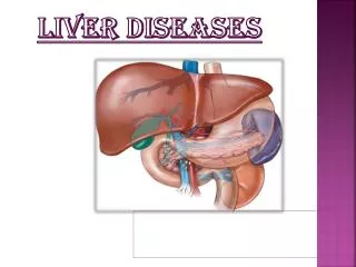
LIVER IN SYSTEMIC DISEASES
LIVER IN SYSTEMIC DISEASES. Dr. Perihan Salem Assistant Professor of Internal Medicine Hepatobiliary Unite. Liver may be involved in wide spectrum of diseases (infections, collagenic, vascular, autoimmune, endocrinal, hematological, malignancies,…..).

LIVER IN SYSTEMIC DISEASES
E N D
Presentation Transcript
LIVER IN SYSTEMIC DISEASES Dr. Perihan Salem Assistant Professor of Internal Medicine Hepatobiliary Unite
Liver may be involved in wide spectrum of diseases (infections, collagenic, vascular, autoimmune, endocrinal, hematological, malignancies,…..). • Liver involvement varies from asymptomatic with abnormal liver biochemistry to florid liver disease.
COLLAGENIC DISEASES: • Systemic lupus erythematosus: * Diagnosis is confirmed by a characteristic autoantibody (ANA, anti-ds DNA). * Liver function test abnormalities are common (~60%), due to: 1- Liver function tests fluctuate with disease activity. 2- Drug therapy (corticosteroids, NSAIDs, immunosuppressive drugs). 3- Activation of occult infection (HBV, HCV). * Findings: • Hepatomegaly. • Abnormal liver function tests (60%) • Liver biopsy: Hepatitis, centrilobular necrosis, nodular regenerative hyperplasia.
Anticardiolipin syndrome: * This syndrome with antiphospholipid antibodies may occur as a primary disorder or associated with other autoimmune disorders (not only SLE). * Among the systemic manifestations as: arterial and venous thromboembolism, recurrent abortions, livedo reticularis (mottled purple discoloration of the skin due to capillary obstruction by thrombi), migraine and thrombocytopenia, the liver may be involved with Budd-Chiari syndrome, focal ischemia, PHT attributed to vascular thrombosis.
Rheumatoid arthritis and Felty’s syndrome: * Abnormal liver function tests (especially ALK.P) mirror disease activity, side effects of drugs and activation of occult infection. * Liver histology shows: fatty infiltration, focal necrosis, nodular regenerative hyperplasia which is related to obliterative vasculitis of small portal veins leading to PHT.
Sjögren’s syndrome: * Primary Sjögren’s (Keratoconjunctivitissicca, xerostomia, salivary gland enlargement) and secondary Sjögren’s (associated with rheumatoid arthritis) are often associated with non-specific changes in liver function tests. * An evident association of Sjögren’s syndrome and chronic HCV infection may represent extrahepatic manifestations of HCV.
Scleroderma: * In the limited form of scleroderma CREST (subcutaneous calcinosis, Reynaud's, esophageal dysmotility, sclerodactyly, telangiectasia), the liver is usually unaffected. * In systemic sclerosis (SS), an overlap with PBC is reported, 25% of patients with scleroderma have +ve AMA, 25% of patients with PBC have anticentromere antibodies of scleroderma and 5-10% of PBC patients have evident scleroderma clinically.
Polyarteritis nodosa and other forms of vasculitis (involving medium/large arteries and hepatic & portal veins): * Abnormal liver function tests during disease activity/side effects of drugs/activation of occult infection. * Hepatic infarction and massive necrosis. * Hepatic venous occlusion and Budd- Chiari syndrome/ PHT.
HEPATIC GRANULOMAS: • Granulomas represent a response to antigenic stimuli. • Causes include: 1- Infection: • Bacterial: Tuberculosis, Brucellosis, Syphilis, Leprosy, Rickettsia (Q fever). • Viral: HAV & HCV (But not HBV), AIDS, Infectious Mononucleosis (CMV). • Fungal: Histoplasmosis (fungal infection: Histoplasma capsulatum), Coccidiomycosis, Blastomycosis. • Protozoa: Toxoplasmosis. • Helminthes: Shistosoma (delayed hypersensitivity reaction against Ag released by the egg), Toxocara Canis (spread by cats and dogs), Fasciola hepatica, Ascaris. 2- Sarcoidosis, PBC, lymphoma, fatty liver. 3- Drug reaction: Granulomas occur as a part of general hypersensitivity reaction developing 10 days-4 months after starting drug intake. Drugs include: carbamazepine; allopurinol; diltiazem; glibenclamide; sulfonamides; quinidine/quinine. 4- Industrial causes: pulmonary & hepatic granulomas may be due to inhalation of cement, mica dust and copper.
Granulomas are of varying size, occuring anywhere in the liver but most frequently near portal tracts. • Granulomas are sharply defined and don’t disturb the normal pattern of the liver. Classically, they consist of pale-staining epithelioid cells with surrounding lymphocytes. Also, giant cells, central caseation and necrosis may be present. Older lesions may be surrounded by fibrous capsule
Specific findings and subtypes of granulomas are: 1- Sarcoid type granulomas: small, well formed and discrete. Multinucleated giant cells and eosinophilic necrosis may be present. Caseation is absent. 2- Caseating (Necrotizing) granulomas: small to large, well formed granulomas with a necrotic center. Common in association with fungal infections, tuberculosis and Hodgkin's lymphoma.
3- Lipogranulomas: composed of aggregates of histiocytes, macrophages and fat. They are usually associated with fatty liver. 4- Microgranulomas: composed of a cluster of 6 or less histiocytes. They have many associations and may represent a non-specific reaction to cell necrosis. 5- Fibrin ring granulomas: They are typical of Q fever, drug reaction (carbamazepine; allopurinol) and acute HAV.
Clinical presentation: • Granulomas are usually asymptomatic. • Palpable liver in only 20% of patients. • Granulomatous hepatitis (usually with infectious causes) is a very rare presentation with active liver disease, marked functional disturbance and liver cell destruction with elevated alkaline phosphatase & transaminases ± fibrosis in liver biopsy.
LIVER IN DM • Type I DM: Hepatomegaly is present in up to 10% of well controlled diabetics and in 60% of uncontrolled diabetics. This enlargement is due to increased glycogen content of the liver. • Type II DM: Hepatomegaly is present due to fatty changes (steatosis, up to 80% of diabetics with fatty liver) which predispose to NASH and steatocirrhosis.
LIVER AND THYROID • The liver plays an important role in the transport, storage, activation and metabolism of thyroid hormones. It synthesizes the proteins which transport thyroxin (T4) in the circulation. The liver stores 10-30% of the body’s exchangeable T4. The liver is the major site for conversion of T4 to its biologically active tri-iodothyronine (T3). The liver removes reverse T3; the biologically inactive product of T4. Also, up to 25% of the daily T4 secreted by the thyroid is metabolized by oxidative deamination/ excreted into bile after glucuronidation & sulphation.
Thyrotoxicosis may induce some abnormalities as: • Slight increase in alkaline phosphatase returning to normal after treatment. • Jaundice in thyrotoxic patient may be due to heart failure and may be without heart failure. • Thyrotoxicosis may aggravate an underlying defect in bilirubin metabolism as Gilbert's syndrome by decreasing bilirubin UDP- glucuronosyl transferase activity.
Myxoedema may induce: * Ascites with or without heart failure, it has been attributed to centrizonal congestion and fibrosis and it disappears on treatment. Ascites shows a high protein content (more than 25 g/L). * Jaundice may occur.
LIVER AND ADRENAL GLAND • Undiagnosed Addison's disease can be associated with mild elevation of transaminases which returns to normal after treatment with corticosteroids.
LIVER AND GROWTH HORMONE • The liver and kidney degrade growth hormone, thus, basal and stimulated growth hormone concentrations are elevated in cirrhotic patients and correlate with the degree of liver dysfunction. • These increased levels contribute to insulin resistance and glucose intolerance in cirrhosis. • Hepatomegaly doesn’t develop despite the chronic elevation of growth hormone in cirrhosis. However, in Acromegaly the liver enlarges in line with other viscera.
LIVER IN AMYLOIDOSIS • Amyloidosis is a condition with extracellular deposition of a protein in an abnormal fibrillar form (AL amyloid, AA amyloid, ATTR amyloid). It is called amyloid because the waxy infiltration of organs resembles starch (Latin: amylum) in its staining. • Clinical features develop because of the disruption of normal function in kidney, heart and other organs by the deposition of amyloid fibrils.
Clinical features: • The common clinical presentation include: nephrotic syndrome, cardiomyopathy, carpal tunnel syndrome, sensory-motor neuropathy, intestinal involvement (motility disturbances & malabsorption) and macroglossia. • Hepatic involvement shows: hepatomegaly, mild elevation of liver enzymes, PHT & liver failure are rare, intrahepatic cholestasis may occur, amyloid fibrils seen by polarization microscopy after Congo red staining.