The History of
180 likes | 383 Vues
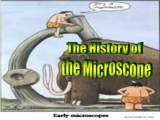
The History of. the Microscope. http://users.adelphia.net/~vtcraig/. A what?!. Microscope:. An instrument for magnifying very small objects. http://inventors.about.com/library/inventors/blmicroscope.htm. In the beginning. The first compound microscope was created by Zacharias Janssen

The History of
E N D
Presentation Transcript
The History of the Microscope http://users.adelphia.net/~vtcraig/
A what?! Microscope: An instrument for magnifying very small objects http://inventors.about.com/library/inventors/blmicroscope.htm
In the beginning... • The first compound microscope was created by Zacharias Janssen • Developed in late sixteenth century • Capable of magnifying images: • approximately three times when fully closed • up to ten times when extended to the maximum http://micro.magnet.fsu.edu/primer/museum/janssen.html
Cells are alive! All cells come from other cells. All living things are made up of cells… Back to Cell Theory... even if it is just one.
Hooke's Microscope What he saw… http://www.mcrit.com/COMSOC/persones_tecniques/Robert_Hooke_archivos/Robert_Hooke.jpg http://cell.sio2.be/introduction/images/cells.jpg
Robert Brown's Microscope http://cip-lx0.physik.uni-siegen.de/~klose/beruehmt/brown_robert_small.jpg http://tiger.towson.edu/~cfanel1/istc301/nucleus.jpg http://www.brianjford.com/wbbrownb.htm
Types of Microscopes • Stereo • Compound light • Brightfield • Darkfield • Phase contrast • Fluorescence • TEM • SEM http://www.louisville.edu/medschool/anatomy/faculty/microscope-cartoon.gif
Stereo Microscopes: • Also called dissecting microscopes • Made of two compound microscopes which focus on the same point from different angles • Low power – cannot see single cells • The specimen is viewed in three dimensions • The image is upright and laterally correct (not upside down and backwards) http://www.microscope-world.com/images/416-microscope.jpg http://www.martinmicroscope.com/MicroscopePages/Stereomicroscopes.htm http://www.umanitoba.ca/Biology/lab1/biolab1_4.html# http://www.ust.hk/~webspade/introduction/Stereomicroscope_eg3.jpg
Compound Light • The most commonly used • Can view individual cells- even living ones! • Image is 2-Dimensional • It has high magnification yet lowresolution • Capable of magnifying images: • 32x • 100x • 400x http://www.southwestschools.org/jsfaculty/Microscopes/types.html http://www.southwestschools.org/jsfaculty/Microscopes/compoundscope.html
1x 32x (Unmagnified) 400x 100x What You See: Elodea http://bio.winona.edu/berg/118s00/Study2/Mvc-006f.jpg http://bio.winona.edu/berg/118s00/Study2/elod32.JPG http://bio.winona.edu/berg/118s00/Study2/elod100.JPG http://bio.winona.edu/berg/118s00/Study2/elod402.JPG http://www.aquaticbiologists.com/images/elodia/elodiaclose.jpg
http://www.olympusmicro.com/primer/anatomy/brightfieldgallery/index.htmlhttp://www.olympusmicro.com/primer/anatomy/brightfieldgallery/index.html Images produced with brightfield illumination appear dark and/or highly colored against a bright, often light gray or white, background. Brightfield http://www.olympusmicro.com/primer/anatomy/brightfieldgallery/bacteriayeastandbloodsmall.html Darkfield Phase Contrast Cell structures appear light on a dark background. Used for looking at living cells. Dense structures appear darker than the background.
http://www.microscopyu.com/articles/fluorescence/fluorescenceintro.htmlhttp://www.microscopyu.com/articles/fluorescence/fluorescenceintro.html Fluorescence • Fluorescence microscopy is used to detect structures, molecules or proteins within the cell • Cell is stained with a dye and the dye is illuminated with filtered light at the absorbing wavelength • Common fluorescence dyes are: • fluorescein~emits green light • Rhodamine~emits deep red fluorescence http://www.ru.nl/molanphys/proteomics.htm http://dept.kent.edu/projects/cell/FLUORO.HTM#Background%20on%20Fluorescence%20Microscopy http://www.microscopyu.com/articles/fluorescence/fluorescenceintro.html http://www.olympusmicro.com/primer/techniques/fluorescence/gallery/cells/header/images/nbl6headersmall.jpg
Optical Electronic vs http://acept.la.asu.edu/PiN/rdg/elmicr/versus.shtml
TEM http://www.udel.edu/Biology/Wags/b617/tem/tem7.gif • Transmission Electron Microscope images are created using electrons instead of light • Electrons travel at shorter wavelengths than light • gives a 2-D view TEM can magnify and resolve a specimen greater than 1,500,000X! • It has high magnification and high resolution http://www.bgsu.edu/departments/biology/facilities/MnM/tem.html http://www.sdsmt.edu/es/emes/tem.jpg
SEM • Scanning Electron Microscopes use electron illumination • The image is seen in 3-D • It has high magnification and high resolution • The specimen is coated in gold • The pictures are in black and white http://www.ptli.com/testlopedia/images/SEM%203%20sm.JPG http://www.uiowa.edu/~image/images/microscopy/EM2/ragweed_pollen_sem.jpeg
Typhoid Mary 1907 http://en.wikipedia.org/wiki/Image:Mallon-Mary_01.jpg#file http://www.todayinsci.com/M/Mallon_Mary/MallonMaryThm.jpg http://www.pbs.org/wgbh/nova/typhoid/mary.html