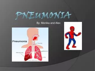
Pneumococcal pneumonia
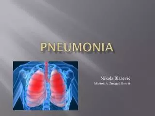
Pneumococcal pneumonia Definition: an acute, suppurative infection of the lungs produced by an encapsulated bacterium, Streptococcus pneumoniae . It is the most commonly occurring bacterial pneumonia in the world. Microbiology encapsulated, Gram ( + ) cocci in chains or pairs

Pneumococcal pneumonia
E N D
Presentation Transcript
Pneumococcal pneumonia Definition: an acute, suppurative infection of the lungs produced by an encapsulated bacterium, Streptococcus pneumoniae. It is the most commonly occurring bacterial pneumonia in the world
Microbiology • encapsulated, Gram (+) cocci in chains • or pairs • does not produce any major toxins, • particularly none that are tissue • destructive
The most important factor defining virulentS. pneumoniae is the presence of a high-molecular-weight complex polysaccharide capsule (Capsular polysaccharide), which is a potent inhibitor of neutrophil phagocytosis
Epidemiology a community-acquired, sporadic disease that occurs most often during the coldest months of the year an occasional cause of nosocomial pneumonia the vast majority of cases occur after aspiration of “normal” oropharyngeal secretions that may contain encapsulated pneumococci, followed by an inability to clear such secretions
Oropharyngeal carrier rates • higher in children (particularly preschool age) than in adults • highest during the coolest months of the year ( when respiratory infections are common) • spread may be enhanced during respiratory tract infections by pneumococcus or certain respiratory viruses such as rhinovirus
Immunology • specific anticapsular humoral antibody (IgM and IgG) • detected in the blood 5 to 10 days after infection • correlates with the clearance of pneumococci and eventual recovery • the major host defense mechanism for eradicating pneumococci: polymorphonuclear leukocytes and alveolar macrophages
Pneumococcal bacteremia • enter the blood stream via lymph channels and the thoracic duct • liver and spleen macrophages rather than polymorphonuclear leukocytes responsible for removing pneumococci from the blood
Congestion“Red hepatization” “Gray hepatization” Resolution 5%~10% patients — pleural space, “empyema” 15%~25% — “bacteremia”
Clinical findings sudden onset — chills, high fever cough: initially — productive of scant mucopurulent or blood-streaked sputum later — thick, purulent, frankly bloody or rust-colored (an alveolar, hemorrhagic, exudative process) pleuritic pain: specific evidence for bacterial pneumonia
Physical examination • Inspection — tachypneic, fever • Signs of consolidation • Pleural effusion • Extrapulmonary infections: meningitis, endocarditis
Laboratory findings WBC :2 to 3 times the normal, bands predominate (left shift ) Chest radiographstypically a lobar distribution with an air bronchogram effect Pleural effusion parapneumonic effusion or empyema Microscopic examination and cultures sputum essential, before therapy is initiated Blood cultures, pleural effusion cultures
Differential diagnosis • Pneumonia caused by different bacteria: microbiologic data • Mycoplasma pneumonia: young patients, prolonged communicability within house-holds. The clinical, radiographic, and pathologic features are usually those of an interstitial pneumonia. Serum cold agglutinin levels may be elevated • Chlamydial pneumonia: the clinical picture is usually that of pharyngitis, laryngitis, and segmental pneumonia of a single lobe without pleural effusion
Legionnaires’ disease • considerable systemic toxicity (fever, chills, myalgia, headache, nausea, vomiting, and diarrhea) • usually occurs in the warmer months or summer • typically male construction workers and smokers in their 50s • dry non-productive cough • non-specific pulmonary infiltrates • Early diagnosis: (1) anti-Legionella fluorescein-labeled antibodies for sputum • (2) antigen detection techniques for urine
Treatment • Antimicrobial therapy • to initiate therapy as promptly as possible • not wait for cultural confirmation • Penicillin G, cephalosporin, erythromycin, clindamycin, or a fluoroquinolone, or vancomycin • initial therapy should be parenteral, be given for at least 5 to 7 days
Supportive therapy bed-rest, monitoring vital signs and urine output, relieve pleuritic pain, replacing fluids, correcting electrolytes, oxygen therapy Complications Empyema: successful antibacterial therapy, drainage ( needle aspiration, chest tubes) Bacteremia: higher dosages of antibiotics, drainage ( septic arthritis, pyogenic pericarditis)
Prognosis case fatality rate untreated: about 25% treated promptly and appropriately:<5%
Prevention • Polyvalent pneumococcal vaccine • healthy adults 65 years or older • those with chronic cardiac or pulmonary diseases, asplenia, chronic liver disease, alcoholism, diabetes mellitus